The Spine, Knees, Ankles & Feet
|

Remember
what happened to the wicked witch? She melted into watery slime. Water doesn’t
work well for structural and mechanical support. Your cells mostly consist of
water. So what maintains their shape? Come
to think of it, your body is also mostly made up water. So what keeps it all together?
What prevents it from falling apart and “melting” into watery slime? What gives
your body’s cells, organs and tissues structural and mechanical support that
lets you keep doing what you need to do to survive and do other things besides?
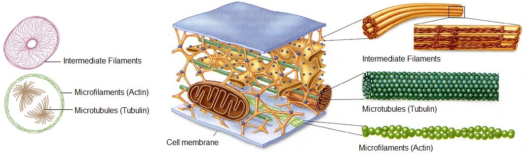
It’s the cytoskeleton of the cell that maintains its shape and gives it structural and mechanical support. It consists of microtubules, microfilaments and intermediate filaments placed at specific sites, made up of specific protein fibers in specific arrangements (Fig.1). Microfilaments provide support below the cell membrane, microtubules crisscross throughout the cell to maintain its shape, and intermediate filaments provide support for the organelles by holding them in place.
Figure 1: The Cytoskeleton |
It’s your connective
tissue that gives your body’s organs and tissues structural and mechanical support.
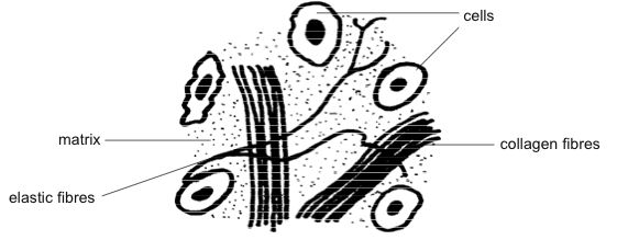
Ordinary connective tissue (Fig. 2) consists of cells called fibroblasts. They
combine water with complex molecules of protein and sugar to produce and send
out a clear, colorless, viscous, gel-like fluid called ground substance
(matrix) which binds all the cells together. Fibroblasts also send out
different types of protein fibers with different mechanical qualities. These
firm collagen, and/or flexible elastin and/or delicate reticular fibers
crisscross through the ground substance to provide the specific organ or tissue
with structural and mechanical support.
The ground
substance and protein fibers make up what is called the extracellular matrix. Bone
and cartilage are called specialized connective tissue due to having
specialized cells that make a specialized extracellular matrix.
Figure
2: Ordinary Connective
Tissue
|
Different
types of connective tissue provide different types of structural and mechanical
support. This runs from firm solid bones, to softer more elastic cartilage, to
high tensile strength ligaments and tendons and to delicate web-like laced spider-like
networks that support most of the organs and passageways in your body. It all depends
on the amount and type of ground substance and the density and mechanical
qualities of the different types of protein fibers or other materials running
through it (like in bone and cartilage).
Ligaments consist of dense fibrous connective tissue. They act like firm ropes to
connect two or more bones together to help stabilize the joint by preventing
the bones from bending, twisting or tearing apart. Tendons, also consisting of
dense fibrous connective tissue, attach muscles across two or more bones
(joints) or to the eye. They need to be sturdy to prevent the muscles from detaching
from the bones.
The cells of cartilage are called chrondrocytes. They produce extracellular matrix that contains chondroitin sulfate (C13H21NO15S) interspersed with different amounts and types of collagen and elastin. This results in various types of cartilage with different qualities, some being sturdier while others more flexible. Besides covering the ends of the bones in your joints (where it is called articular cartilage) it’s also found in the ear, nose, larynx, trachea and large bronchi. It provides support for the respiratory tract, reduces friction within the joints and acts as a shock-absorber for weight bearing joints in the spine and lower extremities.
The main bone
cell involved in its formation and growth is the osteoblast. It lays down a ground
substance called osteoid, a firm organic mesh which is mainly made up of
protein (collagen). It then mineralizes the osteoid by depositing calcium phosphate crystals into it. This
is what gives them the strength to provide support and protection from external
injury for many important organs. The skull protects your brain, the vertebrae
protect your spinal cord, and your ribs and breast bone protect your heart and
lungs.
Your body has over 200 bones, most of which are
tipped with articular cartilage at both ends. Each is precisely shaped with
specific dimensions and comes together with one or more others to form about
360 joints fastened together by over 900 ligaments. Your bones provide attachment
for a few thousand tendons from over 650 skeletal muscles that span across them.
Contraction of these muscles allows you to breathe, open, close and move your
eyes, open your mouth, bite down, chew and swallow and stand up, move around
and handle things.
|
The
Critics Speak
As a result of our upright, bipedal posture, we suffer a huge catalog of woes, including slipped discs, fallen arches and wrenched knees. No engineer, given the opportunity to design human beings from the ground up, would ever dream of confecting a jury-rigged body plan such as ours. But our innumerable afflictions can be understood as the consequence of adapting an ancestral four-legged body to a new bipedal lifestyle.
(Becoming Human: Evolution and Human Uniqueness: Ian Tattersal: New York: Harcourt Brace 1998)
Evolution from walking on all fours to the modern bipedal locomotion is what causes many problems from knee and ankle trouble to lower back pain. To an evolutionary biologist, the appearance of poor design is evidence of the operation of a bungling, unintelligent trial-and-error evolutionary process that has resulted in suboptimal anatomical structures. Biologists point to these sorts of examples because they seem hard to account for if the intelligent design was due to an all-knowing, all-god, all-powerful designer, supernatural or otherwise.
(God, the Devil, and Darwin: Niall Shanks: New York: Oxford University Press: 2004)
Lower back fatigue is mild compared to other problems that are caused directly by design flaws. As our ancestors adapted to a more upright posture, the lumbar area of the vertebral column became sharply curved. The disks of cartilage between each vertebra are not optimally placed for this upright, curved posture; as a result, they sometimes “slip” leading to a painful herniated disk.
The anterior cruciate ligament (in the knee) is vulnerable to tearing because our upright, bipedal posture forces it to endure much more strain than it is designed to. In quadrupeds, the strain of running and jumping is spread among four limbs, and the limb muscles absorb most of it. Once our ancestors transitioned to bipedalism, however, the strain was spread over two legs instead of four. This was too much for the muscles by themselves, so our bodies recruited the leg bones to help with the strain. When you’re running and then stop short or when you make a sharp turn at high speed, the knees must bear the force of this sudden, intense strain. Sometimes the anterior cruciate ligament is simply not strong enough to hold the leg bones together as they twist or pull away from each other.
The ankle contains seven bones, most of them pointless. Because many of the bones of the ankle do not move relative to one another, they would function better as a single fused structure, their ligaments replaced with solid bone. Thus simplified, the ankle would be much stronger, and many of their current points of potential strain would be eliminated. There is a reason that twisted and sprained ankles are so common: the skeletal design of the ankle is a hodgepodge of parts that can do nothing except malfunction.
(Human Errors: Nathan H. Lents; Houghton Mifflin Harcourt: 2018)
Filling
in the Gaps
The Spine
First things
first—the primary purpose of the spinal column is not to let you stand up so you can go where you want to go and
twist this way or that so you can do what you want to do. Although the ease of
spinal movement and its ability to help hold up the body’s weight is very important
for survival, it is secondary to its main purpose of providing a bony canal to
house and protect the spinal cord.
The spinal
cord is part of the central nervous system. It receives and sends sensory
information from throughout your body to your brain so it can know what’s going
on within and around you. It also receives and sends out messages from your
brain to your muscles which signal them to contract in a coordinated fashion so
you can breathe, move around and handle things.
It’s
important to know that nervous tissue is very fragile. With injury or prolonged
pressure nerve cells malfunction and can even die. Now, imagine what would
happen to the unprotected spinal cord of a fetus moving through the birth canal.
An exposed cervical spinal cord under pressure from the narrow but solid tissues
along with twisting the head during birth would likely cause injury resulting
in malfunction. And guess where the messages from the spinal cord come off to
your muscles of respiration to tell you to breathe?
Bingo! They
come from the spinal cord at the 3rd to 5th cervical
vertebrae and go out along the phrenic nerve. Any significant injury to this
region would likely result in apnea—not being able to breathe! This is why a
fracture of the upper cervical spine often results in immediate death.
Do you get the point? Even if the newborn were able to survive delivery, without the spinal cord having solid protection the risk of not being able to breathe properly or even move or feel the arms and legs due to injury of the thoracic and lumbar spinal cord would always be present —not a good formula for survival and being able to reproduce. Let’s take a closer look.
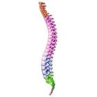
Figure
3: The Spinal (Vertebral) Column
|
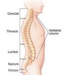
Your spinal
column (Fig. 3) basically connects your head to your pelvis. It consists of
twenty-four separate vertebrae stacked on top of each other—seven cervical
(neck)—twelve thoracic (upper back)—five lumbar (lower back)—followed by the
sacrum, which is attached to your pelvis, and the coccyx (tailbone).
Figure 4: Cross-sectional view of a Vertebra |
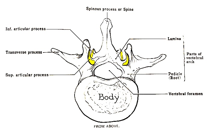
A vertebra
consists of many different parts which together accomplish several functions
(Fig.4). The body sits at the front to provide weight support. The vertebral
arch extends from behind the body, forming the vertebral foramen (where the
spinal cord sits for protection), and consists of the pedicles (extending from
the body) united at the back by the laminae. Three processes—two “transverse”
and one “spinous”—project from the vertebra affording muscle attachment and so act
as levers for neck and back movement. Four articular processes with facets—two “superior”
and two “inferior”—extend up and down from the vertebral arch on either side
forming a facet joint with the corresponding ones on the above and below vertebrae.
They prevent the vertebra from slipping forward, determine the directions of
movement and to what degree, and, depending on the body’s position, sometimes
bear weight. But that’s not all!
Figure 5: Spinal Column and More
|
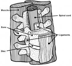
As Figure 5 shows, in between the vertebrae, preventing friction from them rubbing together while providing cushioning that absorbs weight and pressure are the intervertebral discs which are made up of fibro-cartilage. Holding the vertebrae together so they don’t move around and fall apart are numerous ligaments that include those extending up the front and back of the bodies, between the bones making up the vertebral arch and between the transverse and spinous processes. There are also large swaths of dense connective tissue called fascia along with numerous deep, intermediate and superficial muscles attached to the vertebrae by tendons that allow for neck and back support and movement in all directions.
Figure 6: Relationship of Nerve Roots to Spinal Anatomy |
Finally, how
the spinal cord receives and sends out its nerve transmissions is important for
understanding and appreciating the anatomy of the spinal column. The sensory
information the spinal cord sends up to the brain, and the instructions the
brain sends down from to the muscles, enters and exits respectively through the
nerve roots (fig. 6). Space between the
intervertebral disc and notches on the top and bottom of the pedicles form a passageway
between the vertebrae called the neural foramen. Through them pass the
thirty-one nerve root pairs (right/left) that exit the spinal cord; cervical:
8, thoracic:12, lumbar: 5, sacral: 5 and coccygeal:1.
The nerve
roots branch out to eventually form the peripheral nervous system which
connects the spinal cord and brain (central nervous system) to all the sensory
organs and muscles of your body. Each nerve root controls a particular segment
of the body—the lower cervical nerve roots receive sensory data from and send
out motor instructions to the arms—the lower lumbar nerve roots do the same for
the legs. One can therefore see that an injury to the spinal cord and/or a nerve
root because of problems with the vertebral column can result in back and/or
limb pain in addition to partial or complete loss of sensation and motor function
including bowel and bladder control.
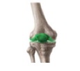
The Knee
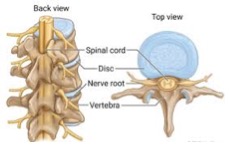
Figure 7: The Knee |
The knee is the largest joint in the body. It plays an important role in bearing your body’s weight and allowing you to be able to walk, run and jump. As Figure 7 above shows, the bones that make up the knee consist of the femur (thigh bone) from above and the tibia (shinbone) from below and the patella (knee cap) in front. The femur ends in two rounded protuberances called condyles which are covered with articular cartilage. They fit nicely into the two grooves at the top of the tibia. In between them providing cushioning and support and reducing friction are the C-shaped lateral (outside) and medial (inside) menisci (singular: meniscus) which are made of cartilage.
The bones of the knee joint are held in place by several ligaments. The anterior cruciate (ACL) and posterior cruciate ligaments prevent the femoral condyles from sliding or rolling backwards or forwards too much on the grooves of the tibia. The lateral and medial ligaments prevent them from moving from side to side too much. When the large muscles attached to the femur and tibia contract they mostly make the knee flex or extend like a hinge but with some degree of rotation. In full extension the knee locks in place thereby resting the surrounding muscles. One can therefore see that an injury or defect in the knee can result in pain along with the inability to stand and move around well.
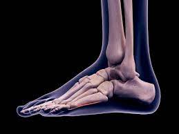
The Ankle/Foot
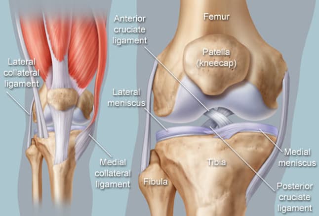
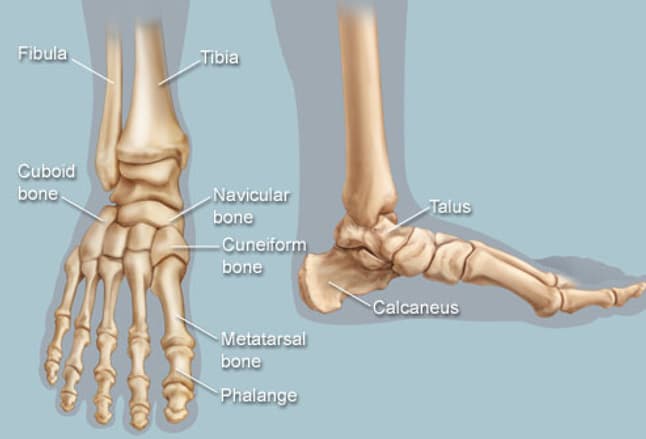
Figure 8: The Ankle/Foot |
The ankle/foot is a complex system made up of 28 bones. In the upright position it supports the body’s weight and like a lever allows us to walk, run, and jump while maintaining our balance. The arch of the foot, which runs from the heel to the ball, provides enough strong and flexible support to afford us these actions (Fig. 8).
As Figure 8 shows, the ends of the tibia and fibula connect with the talus (ankle bone) which sits on the calcaneus (heel bone). The latter two bones form the hindfoot that supports the weight of the body. The midfoot consists of the navicular bone which sits in front of the calcaneus along with four other bones—the cuboid and three cuneiform bones—which sit in front of the navicular bone. They in turn are connected to the five metatarsal bones which begin the forefoot and together with the midfoot form the arch for support, flexibility and shock absorption during ambulation. The metatarsals connect with the rest of the forefoot which consists of the phalanges (digits/toes)—two for the great toe—three for the other four toes. But that’s not all!
|
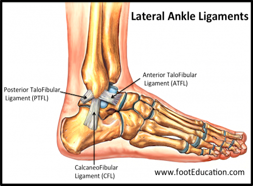
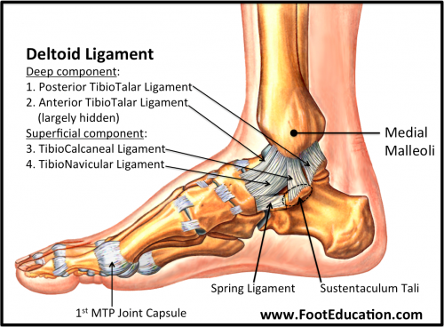
There are 33 joints in the ankle/foot complex each with
articular cartilage for shock absorption and reduced friction. They are held
together by numerous ligaments (Fig. 9) including the ones on either side of
the ankle, within the foot and the plantar fascia that runs almost the entire
length of the bottom of the foot. These joints are moved by over 30 different
muscles with attachments for their tendons, in particular the Achilles tendon,
down the back of the heel which allows for plantarflexion.
The tibia/fibula sit on the talus and act as a hinge joint to afford bending the ankle/foot up and down (dorsiflexion/plantarflexion). The talus on the calcaneus acts as a gliding joint to afford twisting (rotating) the ankle/ foot in or out (inversion/eversion). The metatarsals and phalanges have condyloid joints which afford them bending up or down, sideward movement in or out and circular rotation. All of these play a major role in being able to walk, run and jump while maintaining your balance. One can therefore see that an injury to the ankle/foot can result in pain along with the inability to stand and move around well.
Three Questions
So, besides
how the cytoskeleton that gives structural and mechanical support to your cells
and the different types of connective tissue in your body came to be exactly
where they need to be, there’s a lot more to standing up, walking, running and
jumping while remaining balanced—actions that our spine, knees and ankles and
feet afford us—than what’s mentioned above by these three critics. Here are three
questions to which they don’t even allude, never mind try to answer, which
should give the reader pause.
1.
In what order and from where did the new genetic and
other information come that specifies the size, shape, position, material
specifications and placement of all the parts of the cytoskeleton that affords
our cells structural and mechanical support and what is the real probability
that such a system could have come about by undirected forces while remaining
functional in intermediate organisms each step along the way?
2.
In what order and from where did the new genetic and
other information come that specifies the size, shape, position, material
specifications and placement of all the parts of all the different ordinary and
specialized connective tissues that affords the organs and tissues of our body
structural and mechanical support and what is the real probability that such a
system could have come about by undirected forces while remaining functional in
intermediate organisms each step along the way?
3.
In what order and from where did the new genetic and
other information come that specifies the size, shape, position, material
specifications and placement of all the parts of the spine, knees, ankles and
feet that affords our body the ability to walk, run and jump along with stand
on our toes and stay balanced and what is the real probability that such a
system could have come about by undirected forces while remaining functional in
intermediate organisms each step along the way?
So, are you willing to accept the undirected forces of natural selection acting on random variation as the definitive answer to the above questions? The “smoke and mirrors” of neo-Darwinism which doesn’t even try to account for how each of the parts needed to produce the cytoskeleton of the cells, all the different connective tissues in your body and the spine, knees, ankles and feet came together and happened to have the right size and shape, be in the right position, with the right material specifications for survival?
It’s
important to realize that natural
selection acting on random variation
(genetic mutation) means exactly what it says. Over time, life required a
gazillion bits of new genetic information (not natural selection) to bring
about new structures with new functions. All natural selection did was preserve
the life that was up and running properly and able to survive due to these
gazillion undirected genetic mutations. But keep in mind, natural selection
cuts both ways.
Based on what we know about how life actually works neo-Darwinism may explain the survival of the fittest but not the arrival of the fittest. That’s because, when it comes to survival, logic tells us that the same power that natural selection had to preserve human life when the cytoskeleton in the cells, all the different connective tissues in the body and the spine, knees, ankles and feet worked properly would have also prevented it from surviving if any one of the parts of these structures were either missing, defective, misplaced or not working well or fast enough.
The known engineering principles needed to bring about the functional capacities of all of these parts that resulted in human survival means that, in principle, not only does Darwin’s theory of gradualism fail, but so do all the other neo-Darwinian attempts to replace it. What do you think?
Comments
The group-think of these three neoDarwinian critics (and most others) is that the spine, knee, ankle and foot problems we develop (usually) in later life are a “consequence of adapting an ancestral four-legged body to a new bipedal lifestyle”. They say that this is evidence for “bad design” as “no engineer, given the opportunity to design human beings from the ground up, would ever dream of confecting a jury-rigged body plan such as ours”. One that consists of “suboptimal anatomical structures” some of which are “a hodgepodge of parts that can do nothing except malfunction”. They then, not surprisingly, conclude, without providing any specifics, that “Evolution from walking on all fours to the modern bipedal locomotion” is what caused these problems.
It is hard to believe that they are referring to all of what’s been described above! It’s important to realize that none of the conditions that these critics claim happened due to “bad design” prevented the survival of humanity. Moreover, it’s likely that given the complexity of all of these structures and the physics and material science involved in the functional capacities they afford, an engineer would look askance at their overly simplistic criticisms supported only by their opinions.
It seems really easy to be an evolutionary biologist and critique intelligent design. First, find a condition from which humans suffer. Then, even if it has nothing to do with survival while at the same time not providing an alternative design that makes sense, claim that due to the presence of this condition the organs or tissues involved represent “bad design”. Then, tell a “just so” story, without any specifics, claiming that these organs and tissues came about through Evolution—the undirected forces of natural selection acting on random variation—as the only explanation for causation. The third critic is a master of this technique. Let’s take a closer look.
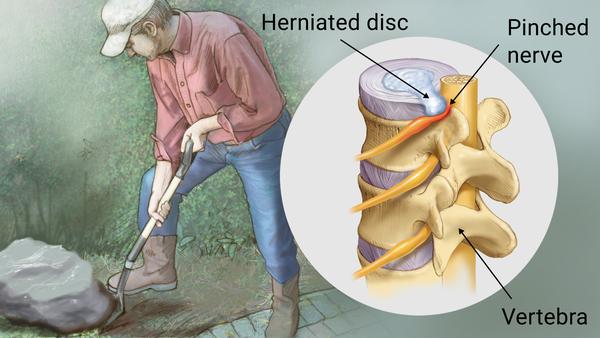
He tells us that “as our ancestors adapted to a more upright posture, the lumbar area of the vertebral column became sharply curved and that the discs of cartilage between each vertebra are not optimally placed for this upright, curved posture; as a result, they sometimes “slip” leading to a painful herniated disk.” (fig.10)
Figure 10: Herniated Lumbar Disc |
The “S-shaped” spine of which the lumbar region represents the lower half is normally not “sharply” but gently curved and along with the gentle outward and inward curves of the thoracic and cervical spines respectively serves to evenly distribute the body’s weight.
What he seems to be talking about regarding the lumbar vertebral disc “not being optimally placed for this upright curved posture” is that (as opposed to a quadruped) when the spinal column is vertical (rather than horizontal) the force of gravity exerted perpendicularly by the vertebra against the disc puts it at risk of slipping out, like toothpaste out of a tube (Fig.10).
When it does this it can often press on
the nerve root nearby and depending on the amount of pressure and how long it
is present, can result in persistent pain down the leg (sciatica) and even loss
of muscle function. This certainly could have impaired the fitness of our ancient
ancestors but since it usually takes place long after their ability to
reproduce would have had zero effect on humanity’s survival.
Yet, given that the way the spinal column is constructed to protect and spinal cord and the placement of the cartilage between the vertebrae to prevent friction and allow flexible mobility one has to wonder where he would propose that it be “optimally” placed? He provides no alternative design that could accomplish the job. To prevent this happenstance in the upright position would require a total revamping of the tissues that provide structural and mechanical support for the spinal cord while allowing for free motion.
It’s easy to critique something when you don’t have to provide an alternative design. He then states the vague neoDarwinian “just so” narrative that “our ancestors adapted to a more upright posture” without any specifics of what this would have practically entailed (see below).
When it comes to the knee he tells us that “the anterior cruciate ligament (ACL) is vulnerable to tearing (Fig. 11) because our upright, bipedal posture forces it to endure much more strain than it is designed to.” Here he seems confused about intelligent design vs the undirected forces of neo-Darwinism as with this statement he clearly agrees that the presence of the ACL in the knee represents design (albeit “bad design”).
Figure 11: ACL tear |
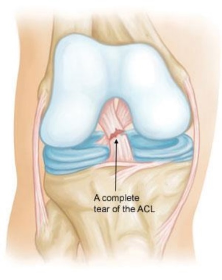
This is an amazing supposition. Since when do limb muscles absorb most of the strain of running and jumping? What’s he talking about and what’s his evidence? Does he have any idea of how the limbs handle the strain of motion? How can he so casually separate out the role that muscles with their tendons play in this situation in contrast to the ligaments? Is he saying that quadrupeds don’t need all of the ligaments in their four limb joints because their muscles handle most of the strain of running and jumping?
The muscles by way of their tendinous attachments to the bones not only cause them to move but also limit bone movement so they do handle some of the strain of running and jumping. But, the position of the muscles affords them only so much strength and they can only react so quickly. That’s why the strong rope-like ligaments positioned exactly where they need to be, connected to each bone within the joint, are there to prevent inadvertent, sudden and excessive hypermobility that may result in partial or full dislocation. Maybe he should speak to people with Ehlers-Danlos Syndrome, a condition of defective connective tissue, to see his assumption and error here.
https://medlineplus.gov/genetics/condition/ehlers-danlos-syndrome/
From his erroneous understanding of how the strain of movement is handled by both bipeds and quadrupeds, he assumes that, on the basis of ACL tears, which almost always take place during athletic related activities that require repetitive jumping, pivoting and rapid changes in direction, (rather than normal everyday human activities), it’s our upright, bipedal mobility that is solely responsible for them. He must not be aware of the fact that most quadrupeds, like dogs, cats, sheep, goats and horses have an ACL in their hind leg knees (stifles). Moreover, large dogs, like Mastiffs, Akitas, St. Bernard's and Rottweilers are prone to ACL tears for the same reason we are—excessive internal rotation of the tibia when the knee joint is partially flexed—which often occurs while running and planting the hind limbs (in humans aggravated by cleats stuck into the ground) while the momentum of the body continues to move forward.
What he seems to miss here is the fact that it is because of the incredible design of our knees, hips and flexible spine with our upright bipedalism that we are able to perform these types of maneuvers which indeed put us at risk for injury (see below). It is certainly possible that in pursuing or being pursued our ancient ancestors could have sustained an ACL injury which would have left them vulnerable. But since humanity has existed for a long time, it would appear that when it comes to our survival this critic’s concern is unfounded.
Regarding the ankle, this critic opines that it “contains seven bones, most of them pointless” (elsewhere he says the same about the eight bones of the wrist). And that since most of them don’t move with respect to each other it would be better to replace their ligaments, which are prone to injury, with bone to make it stronger. He then uses this baseless and bizarre position to support his neoDarwinian claim (of apparent design without a designer) that “the skeletal design of the ankle is a hodgepodge of parts that can do nothing except malfunction.”
This statement alone forces us to question the critic’s knowledge of biomechanical function when it comes to the ankle and foot. The seven bones of the ankle that he’s referring to are the talus, calcaneus, navicular, cuboid and the three cuneiforms. However the ankle also involves the fibula and tibia (fig.8). Although he states that “many of the bones of the ankle don’t move relative to one another”, he doesn’t clarify which ones he’s talking about.
Since we know that the talus moves up and down below the fibula and tibia and glides in and out over the calcaneus we must assume that he’s referring to the navicular, cuboid and three cuneiforms. These perfectly shaped and positioned bones of the midfoot are held together by strong ligaments to form part of the arch of the foot that makes it strong enough to allow for weight bearing but are flexible enough to allow for up and down movement during walking and running. One can immediately see that fusing these specific bones would have a negative impact on ankle and foot function.
However, when it comes to ankle injuries it’s not the ligaments of the midfoot that are usually involved. It’s usually the lateral ligaments that get injured due to a combination of sudden plantarflexion and internal rotation, like when quickly pivoting and changing directions in basketball (see fig.9: note the lateral and medial ligaments of the ankle but also the ligaments holding the bones of the midfoot together). In other words, this critic seems to be mixed up about not only how the ankle works with respect to the foot, the necessity of the strength and flexible nature of the midfoot (the latter would be severely compromised by bony fusion) and which ligaments of the ankle tend to get injured and why.
At the end of the day, he gives us no reasonable mechanism for how this all came into being. Instead, alluding to the magic of Evolution, he tells us that it all consists of “a hodgepodge of parts that can do nothing except malfunction”. Well, it seems to have worked well enough to keep humanity alive and functioning for a long time despite his criticism.
Laufmann’s Triple Filter
Not
understanding the objectives of the designer
Evolutionary biologists seem to be in agreement about the advantages and “reasons” why humanity developed the ability to stand and walk on two legs (upright bipedalism). They include providing energy efficient locomotion that favors slower speed but longer distance, better vision above the grass to allow for better predation and defense and decreasing exposure to the heat of the sun. But the most obvious advantage was that standing erect and moving with alacrity on two legs allowed humans to free up their forelimbs to, among other things, stretch for fruit in trees, carry food and babies, make, carry, and use tools and weapons, and socially integrate with non-verbal communication.
Maybe you’re asking yourself; “Those are really good reasons for why upright bipedalism is important for human survival, but how exactly did it all come into being?”
Anyone who knows anything about how the body works should realize that with all these new and improved human capabilities this would have meant that besides the necessary anatomical changes to achieve efficient upright bipedal mobility so would there have been physiological ones as well. This would have included; with a higher center of gravity being able to run, jump and spin around in all directions while staying balanced on just two feet; in the upright position preventing gravity from reducing enough blood flow to the brain to cause passing out; having the manual dexterity and creative and language capabilities afforded by the neuromuscular system under control of the larger brain to take advantage of upright bipedalism. After all, if we didn’t have any of these capabilities, then what would have been the use of standing up on two feet?
Without providing any understanding or explanation of what it takes for human upright stature and two legged locomotion our second critic dogmatically tells us that what brought it into being was a “bungling, unintelligent trial-and-error evolutionary process”.
Rather than accepting this vague and essentially useless neoDarwinian narrative, it might instead be good to look at what a distinguished professor of mechanical engineer with extensive design experience actually has to say about this topic. While reading what follows ask yourself if evolutionary biologists have the right to tell us about human origins if they don’t even mention, never mind, try to explain, how this could have come about—remember, no magic allowed.
In
his book “The Design and Origin of Man” Professor Stuart Burgess details several
“unique features” along with “unique abilities” present in bipedal humans
compared to our nearest relative, the quadrupedal ape. https://www.youtube.com/watch?v=Yd7nUN-n2c4
He
notes that “according to evolution, humans have gradually evolved an upright
stature over millions of years, (but) to stand upright, humans need many design
features, not present in quadrupeds and these must all be in place
simultaneously.”
In
contrast to the neoDarwinian narrative which totally ignores
these required anatomical and physiological features, Dr. Burgess applies his
extensive engineering experience to explain this literally from the ground up.
In other words, our upright bipedalism and the unique mobility and functional
capabilities it affords us, starts with having the right type of feet and ends
with having the right type of brain to maintain control. Let’s take a closer look.
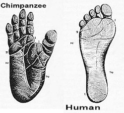
Dr.
Burgess begins by pointing out that the human foot (fig.8) doesn’t have just
one arch running from its heel to its ball, resulting in only two contact
points with the ground. On closer study, the human foot actually has three
arches and three points of contact with the ground. Just like the three legs of
a stool provide it with better stability, Dr. Burgess tells us that “it is well
known in engineering that the most precise way of supporting an object is to
have three points of contact.”
The
three points of contact with the ground for the human foot are the center of
the heel, the ball of the foot near the big toe, and the ball of the foot near
the little toe. And the three arches are the two from the heel to the ball of
the foot near the big toe and near the little toe and the one across the ball
of the foot from these points of contact. It’s the perfectly shaped and positioned
bones of the foot along with its ligaments and tendons that form these arches.
They let us stand on one leg while placing our body’s center of gravity between
these three points of contact.
In
addition, the human foot has a uniquely strong and stiff big toe, close to the
others, that provides enough power for the final push while walking or running.
An ape instead “has very flexible feet that are effectively a second pair of
hands” as its “big toe is a flexible thumb that is designed to grab branches”
(Fig.12). That’s why “when apes attempt
to walk with an upright stature, they cannot make a firm push from their big
toe.” That explains a lot, doesn’t it?
Figure 12: Ape and Human Foot |
Humans
have legs that are longer, being one-half of the total length of the body versus
one-third for apes (Fig. 13). Having longer legs and shorter arms allows us to
easily walk and run while using our upper limbs whereas apes have shorter legs
and longer arms so they can reach the ground to allow for four-legged motion.
Dr. Burgess then points out that in the evolution of a quadruped to a biped,
“having longer legs before being able to stand upright would (interfere with
the arms striking the ground and would) make it difficult to walk, not only on
two but also four legs.”
|
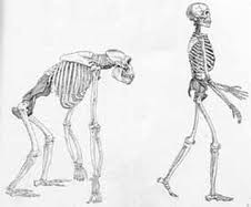
Our uniquely upright knees
that can lock in place, angled inward thigh bones and upright hip joints all
work to let us easily stand up on two legs for a long time and move quickly. Also,
our gently curved “S” shaped spine “is ideal for an upright stature because on
the one hand it produces a straight back but on the other hand it cushions the
spine when it is subject to a compression load.” In fact our straight back is
perfect for an upright stature because “the torso and head are directly above
the hips in the standing position”. In contrast, the bent “C” shaped spine of
the ape “makes their torso project out in front of the hips so they must use
their arms and hands to support their weight, making them “knuckle-walkers””
(Fig.13).
For humans, the spinal cord enters the skull to connect up with the brain through the bottom of the skull (fig. 13). So, for humans, the natural placement of the head allows them to look forward when in the upright position. In fact, when on all fours we have to force our head upwards to keep looking ahead. In contrast, for apes, the spinal cord enters the skull to connect up with the brain through the back of the skull. So, for apes, the natural placement of the head allows them to look forward when in the horizontal position. In fact, when standing on two legs they have to bend their head downwards to keep looking forward. Looking at where the opening in the skull is for the spinal cord is one way that paleontologists can determine if the animal walked on two or four legs. Great, but how did the skull get that way? That’s a lot of anatomy to change!!
Humans have a flatter face than apes (Fig. 13) which with downward eye movement allows them to see the ground on which they are moving easier. For an upright biped, like us, with a higher center of gravity and only two feet in contact with the ground, this is very important for staying balanced as our visual cues play a major role in stabilization especially with a sudden loss of footing. In contrast, apes have a large protruding jaw (Fig. 13) that limits their view of the ground on which they are moving. However, being lower to, and having four limbs in contact with, the ground makes it much easier for them to remain balanced.
To remain upright and balanced while standing, walking, running and jumping your brain, in addition to making sure there’s enough blood flowing to it in the upright position, uses the sensory information from your eyes, vestibular apparatus in your ears, pressure sensors in your feet and position sensors in all of your muscles, tendons and joints to maintain your balance.
It also affords us, as Dr. Burgess says “amazing coordination between the hand and the eye during walking and running” in addition to fine motor dexterity and being able to see up close through accommodation. Without all of these innovations to allow for upright bipedalism what would have been the use of having a brain that could coordinate these actions?
Before,
moving on to the next section review all of the anatomical differences between
a human and an ape that allows for upright bipedal mobility and ask yourself if
the neo-Darwinian explanations given by these critics answers your questions
and satisfies your curiosity.
Not
accounting for the functional requirements, constraints and trade-offs
If
we can agree that the main advantage for human upright bipedal mobility was the
functional requirement of to stand, walk, run and jump while freeing up the forearms
for defense, work, creative activities and non-verbal communication, then the
most obvious constraint resulting in trade-offs would be the force of gravity.
In
the upright position a biped with a higher center of gravity and only two
points of contact with the ground is inherently more unstable than a quadruped
(see above). In other words, the main constraint of upright bipedalism is the
increased risk for falls which can cause life-threatening brain, bone and other
injuries. Falling is very prevalent early in life as toddlers learn to stand
up, walk and run, and again, later in life when the elderly suffer from
arthritis, muscle weakness and imbalance.
It’s
incredible that none of these critics (and others) even mention this obvious risk.
Rather, they point out only later non-life-threatening problems like back pain,
sore knees and fallen arches. Maybe it’s because they’d have to explain how the
human body developed the ability to stay balanced, especially when going from
four-limbed to two-limbed mobility. The continued existence of humanity clearly
shows that having the ability to be mobile on two legs with the balance and coordination
it requires apparently outweighs the risk for life-threatening fall injury.
Failure
to acknowledge user abuse and degradation over time
The
commonest cause for low back pain is muscle strain and soft tissue injury, not
a slipped disc. As people age they become more susceptible to low back pain due
to obesity, lack of exercise, poor posture, improper lifting and degenerative
arthritis in the vertebral joints. Knee abuse in sports, causing ligament and
cartilage injuries, and aging can lead to painful arthritis. Ankle sprains and
Achilles tendon rupture can take place due to user abuse and aging as can
various foot ailments as well. Even the best designed machines are
subject to sometimes not being used properly or degeneration. Nothing lasts
forever.
Conclusion
That our spine, knees, ankles and feet
can cause suffering from fall injuries, user abuse and degenerative changes, is
only made possible by the anatomical and physiological innovations needed to
afford us two-legged mobility. To claim that this represents “bad design” in
the human body is not only misguided but, based on engineering principles,
totally absurd. It would seem that, without accounting for all of what was
necessary to achieve it, the theory that it came into existence by “bungling, unintelligent trial-and-error” actually describes the
theory itself rather than the actual mechanism. What
do you think?
Also see Dr. Glicksman's Series on
"Beyond Irreducible Complexity"
"Exercise Your Wonder"
Howard Glicksman M. D. graduated from the University of Toronto in 1978. He practiced primary care medicine for almost 25 yrs in Oakville, Ontario and Spring Hill, Florida. He now practices palliative medicine for a Hospice organization in his community. He has a special interest in how the ethos of our culture has been influenced by modern science’s understanding and promotion of what it means to be a human being.
Copyright 2022 Dr. Howard Glicksman. All rights reserved. International copyright secured.