|
|||||||||||||||||||||||
|
|||||||||||||||||||||||
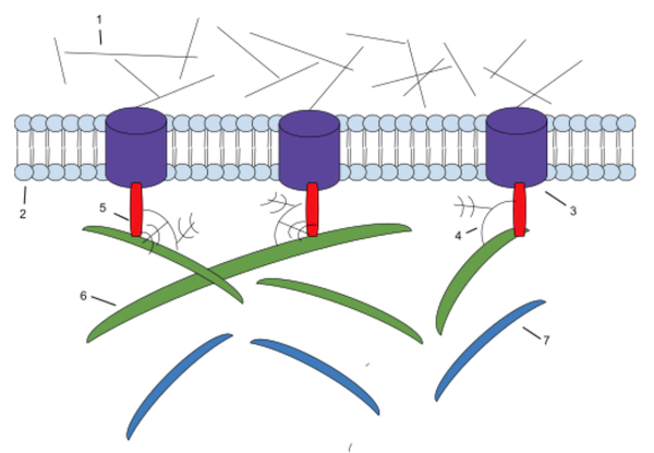
However, your cells mostly consist of water and so do all the tissues and organs in your body. So, how do they maintain their shape and what gives them structural and mechanical support? As a prior article noted it is the cell’s cytoskeleton (microtubules, microfilaments and intermediate filaments) that give it shape and structural and mechanical support. And it is the connective tissue, consisting of cells (mostly fibroblasts) that secrete a gel-like ground substance and protein fibers, that crisscross through it, which provide your body’s tissues and organs with structural and mechanical support. The ground substance and protein fibers are called the extracellular matrix (ECM) which is the non-cellular component of connective tissue. In fact, different types of connective tissue provide different types of support. In the human body this runs from solid bones, to softer and more elastic cartilage, to high tensile strength ligaments and tendons, to the delicate web-like laced spider-like networks (bubble wrap) that supports most of its organs and passageways. It depends on the different types of cells which secrete different types of ground substance and the density and material qualities of the different protein fibers running through it. But the ECM does much more than just provide the body’s tissues and organs with structural and mechanical support. It also affects cell signaling, migration, growth, proliferation, differentiation and survival, all of which regulates tissue morphology, development, homeostasis and function. The ECM manages these functions through its main components, collagen and elastin fibers (see ECS-12), the gel-like ground substance made of water, glycoaminoglycans (GAGs) and proteoglycans (PGs - see ECS-13), glycoproteins (GPs), and receptors like integrins, (see ECS-14), growth factors (GFs), enzymes and more (Fig.1).
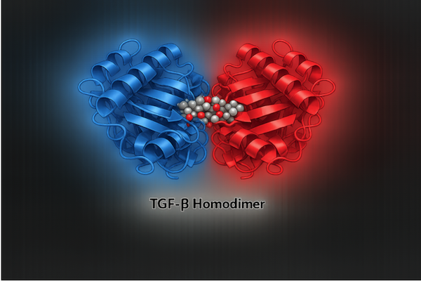
This article will look at GFs, cytokines, chemokines, matrikines, enzymes and their inhibitors. Whereas the three prior articles surveyed all the structures of the ECM, this one will review how these six important factors work to maintain control of ECM structure and function. In general, GFs define and regulate the high-level tissue program; cytokines modify that program by imposing the immune context that governs inflammation, defense, and repair; chemokines further refine it by directing cell movement; matrikines report the mechanical and biochemical state of the ECM and trigger the corresponding cellular response to that state; enzymes remodel the ECM and generate matrikines; and enzyme inhibitors apply the braking force that stabilizes remodeling and prevents runaway degradation. Keep in mind, that we are supposed to believe that an unguided process, like natural selection acting on random variation, was responsible for the presence of each of these components and what they, in combination, do for the body. Growth Factors (GFs) There are about 300 genes that encode for the several different ECM-associated GF families and their specific receptors. This includes the TGF-β family (Transforming Growth Factor-beta), VEGFs (Vascular Endothelial Growth Factors) and PDGFs (Platelet-Derived Growth Factors) which this article will review. Any cell that builds, remodels, or responds to the ECM can produce GFs. This includes fibroblasts, macrophages, platelets, epithelial cells, endothelial cells, smooth muscle cells, chondrocytes, osteoblasts, and many embryonic cell types. GF production is controlled by transcription factors, mechanical forces, cytokines and inflammatory signals, cell–cell contact, ECM stiffness and composition, metabolic state, injury-associated cues such as hypoxia or tissue damage, and developmental patterning systems. These controls operate in adult tissues with continuous turnover (such as skin and intestine), in wound healing, tissue repair, and embryonic development. After GFs are secreted by cells, they generally end up in one of three locations. A minority remain freely soluble in the interstitial fluid until they are captured by cell-surface receptors or by the ECM. When they bind to the ECM, they can form gradients and long-term storage pools for later signaling. Some are tethered to the cell-surface or the local ECM staying there until released by specific enzymes or mechanical stress. Many attach to structural ECM proteins such as collagen, fibronectin, laminins, and, in particular, heparan sulfate proteoglycans (HSPGs), like perlecan, in the basement membranes, to be later released by enzymes or integrin-associated mechanical tension. Transforming Growth Factor-beta (TGF-β) When injury occurs, this same system is redirected into repair mode. TGF‑β becomes one of the key chemical signals that organizes wound healing by shifting the post‑injury program from breakdown and inflammation into tissue rebuilding and scar formation. It recruits fibroblasts into the damaged area and regulates the production of ECM components such as collagen, fibronectin, laminins, proteoglycans, and related matrix proteins. At the same time, it decreases ECM breakdown by suppressing matrix‑degrading enzymes and increasing production of their inhibitors, keeping the rebuilding phase structurally controlled and directed. TGF‑β’s logic originates in development: in the embryo it drives broad cell‑fate decisions and mechanically organizes tissues by controlling how cells differentiate, position themselves, and assemble the structural matrix. In adult tissues, this same program is repurposed to maintain equilibrium by suppressing unnecessary proliferation, limiting immune activation, and keeping ECM production and degradation in balance. In this setting, TGF‑β functions as a brake that restrains growth, quiets immune activity, and stabilizes tissue architecture. When cancer emerges, malignant cells selectively disable the growth‑inhibitory arm of the pathway while continuing to exploit its immunosuppressive and matrix‑modifying outputs. The homeostatic brake is lost, but the pro‑invasion and pro‑tolerance functions remain intact, allowing tumors to proliferate unchecked while shaping a permissive microenvironment. Cells normally increase TGF‑β production when they encounter conditions that signal mechanical strain, injury, or threat. These include mechanical stress transmitted through integrins, inflammatory cytokines released by macrophages and damaged cells, chemicals released from platelets and clotting factors during tissue injury, low oxygen tension, pathogen‑associated signals during infection, and ECM degradation fragments generated by enzymes (matrikines). These inputs activate the same system that maintains homeostasis, allowing it to respond proportionally to mechanical load, inflammation, or damage. TGF‑β contains two identical protein chains of 110 amino acids joined by disulfide bonds (Fig.2).
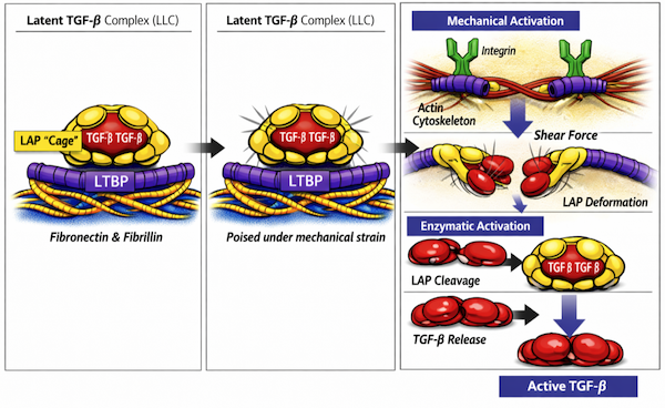
TGF-β is sent into the ECF in an inactive, or latent, form. It is synthesized in the endoplasmic reticulum and wrapped within a protective “cage” called the LAP (latency-associated peptide) which inactivates it by physically blocking its receptor-binding sites. The LAP is then linked to the LTBP (latent TGF-β binding protein) and this large latent complex (LLC) is sent out into the ECF, where it tethers itself to glycoproteins like fibronectin and fibrillin (Fig.3). Latent TGF-β becomes activated when the LAP cage is either physically deformed or enzymatically broken apart. Physical deformation occurs when increased tissue stretching, tension, or remodeling is sensed by integrins on the cell surface, which are linked to the cytoskeleton and transmit pulling forces that distort the LAP. Enzymatic activation occurs when injury, inflammation, coagulation, or wound-healing signals trigger the release of specific proteases that cleave the LAP (Fig.3).
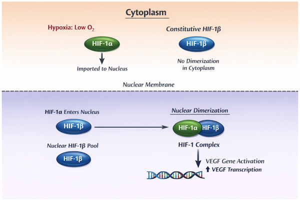
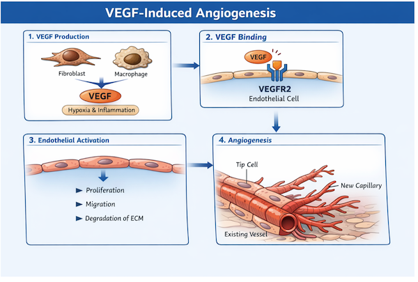
Once TGF-β is freed from the LAP “cage”, its receptor-binding sites become exposed and it can attach to TGF-β Receptor Type II on the cell surface. This initiates a signaling cascade involving multiple intracellular proteins and hundreds of transcription factors, activating and repressing large gene sets to produce the many above-mentioned functions attributed to TGF-β. Vascular Endothelial Growth Factors (VEGFs) Hypoxia is one of the main initiating signals that drives VEGF secretion in tissues that lack adequate oxygen delivery. All cells produce Hypoxia-Inducible Factors 1α and 1β (HIF-1α and HIF-1β) within the cytoplasm. In the presence of adequate levels of oxygen, HIF-1α is readily destroyed by a series of oxygen-dependent enzymes. When there isn’t enough oxygen present, this allows HIF-1α to accumulate in the cytoplasm and through an active import mechanism, move into the nucleus. In contrast, under normal circumstances HIF-1β is constitutively formed within the cytoplasm and, since it is not targeted for enzymatic breakdown, remains stable and continuously enters the nucleus. Once inside the nucleus, HIF-1α joins with the pre-existing pool of HIF-1β, to form a transcription-factor complex that activates VEGF gene expression (Fig. 4).
There are several isoforms of VEGF but the dominant one is VEGF165 which is a homodimer of two identical polypeptide chains containing 165 amino acids joined together by disulfide bonds. After secretion, some of it remains soluble and freely diffuses through the interstitial fluid, but most of it binds to HSPGs, like perlecan, and other ECM glycoproteins, such as fibronectin and tenascin-C. This bound VEGF can be released only by specific enzymes that break these connections which are induced during acute inflammation, tissue injury and repair, hypoxia, and active ECM remodeling. VEGF triggers angiogenesis, the process in which endothelial cells open a path by degrading the ECM, then sprout, migrate, and proliferate from an existing vessel to form new branches (Fig. 5).
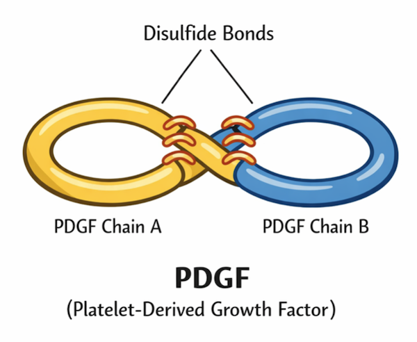
VEGF binds to its specific receptor, VEGFR2 (Vascular Endothelial Growth Factor Receptor 2) which is expressed mainly on endothelial cells that line all blood vessels. This receptor activation shifts endothelial cells from a resting state to an active growth-and-movement state by turning on intracellular signaling programs that alter transcription factor activity and reprogram the cells for the behaviors required to build a new sprouting blood vessel. Platelet-Derived Growth Factors (PDGFs) There are four different gene‑encoded PDGFs. PDGF‑A has about 200 amino acids, PDGF‑B about 240 amino acids, and PDGF‑C and PDGF‑D about 350 amino acids each. They are joined together by disulfide bonds, and assemble as specific dimers—AA, AB (Fig.6), BB, CC, and DD. PDGF‑AA, AB, and BB are secreted in active form, whereas PDGF‑CC and PDGF‑DD are secreted as inactive precursors that bind to HSPGs in the ECM requiring enzymatic cutting to activate them.
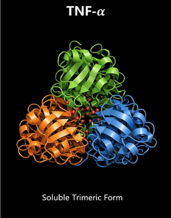
Platelet-derived growth factor receptor alpha (PDGFR-α) and platelet-derived growth factor receptor beta (PDGFR-β) are the two receptors that bind PDGF dimers. The cells with these receptors are mainly fibroblasts, connective-tissue progenitor cells, pericytes, and vascular smooth muscle cells. When a PDGF dimer binds its receptor, this triggers intracellular signaling systems that activate and repress transcription factors to turn many genes on or off, controlling cell proliferation, migration, matrix production, and vessel stabilization. Together, the PDGF system coordinates mesenchymal responses that drive tissue repair and angiogenesis. Cytokines There are about 100 genes that encode for inflammation-related cytokines and their receptors. This includes Tumor Necrosis Factor (TNF), Interleukins (ILs), and Interferons (IFNs). They are produced by immune cells such as macrophages, neutrophils, dendritic cells, NK (natural killer) cells, T cells, and B cells, and by non-immune structural cells such as fibroblasts, pericytes, endothelial cells, epithelial cells, adipocytes, and smooth muscle cells. Their release is triggered by immune cell activation, infection, tissue damage, metabolic stress, mechanical stress, and other cytokines. After release, cytokines bind specific receptors on target cells and activate signaling pathways that drive inflammation’s core processes: immune cell recruitment, vasodilation, metabolic reprogramming, proliferation, wound healing, tissue repair, and survival. Tissue Necrosis Factor-alpha (TNF-α)
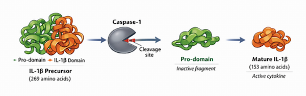
TNF-α binds two receptors—TNFR1 and TNFR2—which are expressed on endothelial cells, fibroblasts, epithelial cells, smooth muscle cells, macrophages, dendritic cells, and lymphocytes. Binding these receptors activates intracellular signaling pathways that amplify inflammation and coordinate the tissue response. A major outcome is white blood cell recruitment: endothelial cells increase their surface adhesion molecules and also become more permeable, allowing circulating leukocytes to adhere to and migrate between them into the ECM. Additional receptor-driven responses across immune and structural cells promote pathogen control, limit the spread of tissue injury, and initiate early steps of tissue repair. Interleukin-1beta (IL-1β) IL-1β is first synthesized as an inactive precursor, consisting of 269 amino acids, that is stored in the cytoplasm. Inflammation generates chemical “danger” signals that activate caspase1, an enzyme present in an inactive form inside the cytoplasm of macrophages and dendritic cells. Once activated, caspase1 cleaves and activates the stored IL-1β precursor in the cytoplasm, allowing the mature cytokine, containing 153 amino acids, to be released into the ECM where it increases the inflammatory response (Fig.8).
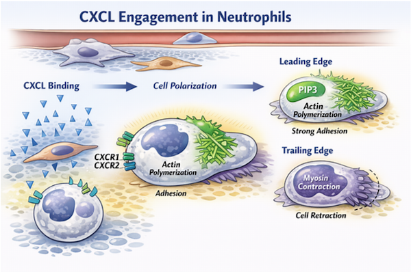
IL-1β exerts its effects by binding to the IL-1 receptor type 1 (IL-1R1), present on endothelial cells, fibroblasts, epithelial cells, smooth muscle cells, macrophages, dendritic cells, and lymphocytes. Receptor engagement activates intracellular signaling pathways that amplify inflammation and coordinate the tissue response. A major outcome is leukocyte recruitment: endothelial cells increase adhesion molecules and increase permeability, enabling circulating white blood cells to adhere and migrate into the ECM. Additional receptor-driven responses across immune and structural cells enhance pathogen control, expand local inflammation, and initiate early steps of tissue repair. Interleukin-6 (IL-6) IL-6 is a multifunctional proinflammatory cytokine containing 185 amino acids that is released by macrophages, dendritic cells, neutrophils, T cells, B cells, fibroblasts, endothelial cells, fat cells, and skeletal muscle cells. These cells secrete IL-6 when they detect infection, tissue damage, metabolic stress, or when activated by inflammatory cytokines such as TNF-α and IL-1β. IL-6 is released in an active form. It binds to the IL-6 receptor complex on liver cells, endothelial cells, fibroblasts, epithelial cells, macrophages, dendritic cells, and lymphocytes. Once engaged to the receptor, IL-6 activates intracellular signaling pathways that coordinate both local and systemic inflammation. A major outcome is induction of specialized proteins by liver cells, which enhance pathogen control and modulate inflammation. Additional receptordriven responses support leukocyte recruitment, promote lymphocyte survival and differentiation, and initiate early steps of tissue repair. Interleukin-10 (IL-10) IL-10 is an important anti-inflammatory cytokine containing 178 amino acids that is released primarily by regulatory T cells and macrophages, but also by dendritic cells, B cells, NK cells, epithelial cells, and fibroblasts. These cells secrete IL-10 in response to ongoing inflammation, immune activation, and cytokine stimulation, such as TNF-α, IL-1β, IL-6, and IFN-γ — which act as a built-in brake to prevent runaway inflammation. IL-10 is released in an active form. It acts by binding to the IL-10 receptor on macrophages, dendritic cells, lymphocytes, endothelial cells, and fibroblasts. Receptor engagement activates intracellular signaling pathways that suppress inflammation and restore tissue homeostasis. A major outcome is inhibition of macrophage and dendritic cell production of TNF-α, IL-1β, and IL-6, thereby limiting further leukocyte recruitment. Additional receptor-driven responses promote resolution of inflammation, support tissue repair, and maintain epithelial and stromal integrity. Interferon-gamma (IFN-γ) IFN-γ is a proinflammatory cytokine containing 189 amino acids that is released primarily by NK cells and different types of T cells. They secrete IFN-γ when they detect intracellular pathogens, receive antigen-driven activation signals, or are stimulated by cytokines such as IL-12 and IL-18. IFN-γ is released in an active form. It acts by binding to the IFN-γ receptor on macrophages, dendritic cells, endothelial cells, fibroblasts, epithelial cells, and lymphocytes. Once engaged to the receptor, IFN-γ activates intracellular signaling pathways that strengthen antimicrobial defense and coordinate cellmediated immunity. A major outcome is macrophage activation: macrophages increase antigen presentation, microbicidal activity, and production of reactive oxygen and nitrogen species. Additional receptordriven responses enhance leukocyte recruitment, reinforce some T cell immunity, and promote containment of intracellular pathogens. Chemokines Chemokines are small secreted proteins of about 70–90 amino acids that direct cell migration (chemotaxis) along concentration gradients. Their folds are stabilized by cysteine residues, and their nomenclature reflects the spacing of these cysteines. CXCL chemokines contain two cysteines separated by one amino acid (C-X-C), and CCL chemokines contain two adjacent cysteines (C-C). The “L” indicates a ligand. Across the genome there are roughly 50 chemokine and chemokine-receptor genes, including CXCL1, CXCL2, and CXCL8, which signal through seven CXCRs, and CCL2, CCL3, CCL4, and CCL5, which signal through ten CCRs. The CXCL family of chemokines is predominantly secreted by epithelial cells, endothelial cells, macrophages, and fibroblasts. These cells respond to biochemical markers of acute infection (PAMPs = pathogen-associated molecular patterns), to cytokines such as TNF-α, IL-1β, and IFN-γ, and to tissue-injury signals including hypoxia, metabolic stress, and mechanical stress. CXCL chemokines rapidly recruit neutrophils to tissues in the early phases of inflammation. In contrast, the CCL family of chemokines is predominantly secreted by macrophages, dendritic cells, fibroblasts, and endothelial cells. Their release is driven mainly by cytokines such as IL-1β, TNF-α, and IFN-γ, and by local inflammatory signals, with more limited stimulation by PAMPs. CCL chemokines recruit monocytes and macrophages and become more prominent in the later phases of inflammation, when tissue repair and remodeling begin. Differential CXCL vs CCL function—and therefore neutrophil vs monocyte/macrophage migration—arises from three linked mechanisms: (1) stimulus-specific chemokine release (2) differential chemokine immobilization on GAGs, and (3) differential receptor density and signaling on the responding leukocytes. Local release of CXCL or CCL chemokines by epithelial cells, endothelial cells, fibroblasts, and macrophages occurs only when the appropriate stimulus is present. Once secreted, CXCLs and CCLs diffuse outward into the ECM. Their diffusion is immediately shaped by their distinct affinities for GAGs such as heparan sulfate, chondroitin sulfate, and dermatan sulfate on PGs displayed by endothelial cells, fibroblasts, epithelial cells, and the ECM itself. This selective GAG binding converts a broad cloud of chemokines into a stable, spatially precise gradient. Neutrophils express a high density of CXCR1/2 and comparatively low CCR expression. In the early inflammatory phase—when CXCL1, CXCL2, and CXCL8 dominate—neutrophils bind CXCLs strongly and efficiently. CXCR binding polarizes the cell, which means that it becomes physically asymmetric and forms a leading edge and a trailing edge. This occurs because CXCR signaling reorganizes the cytoskeleton and adhesion machinery in a directional way: CXCR engagement produces a localized intracellular signal (PIP3) at the future leading edge, which initiates actin polymerization and establishes polarity, adhesion increases at the front, and the rear contracts. The cell then crawls up the CXCL gradient (Fig.9).
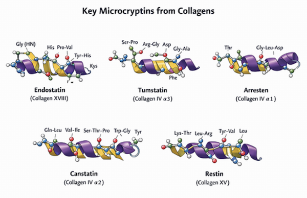
Monocytes and macrophages express a high density of CCR1/2/5 and comparatively low CXCR expression. In the later inflammatory phase—when CCL2, CCL3, CCL4, and CCL5 dominate—monocytes and macrophages bind CCLs strongly and efficiently. CCR engagement triggers the same polarity program: a localized intracellular signal (PIP3) forms at the leading edge, actin assembles at the front, the rear contracts, and the cell moves persistently toward the higher CCL concentration. Matrikines Enzymatic cleavage produces matrikines by cutting peptide bonds and releasing free, soluble fragments, whereas mechanical unfolding produces them by exposing hidden signaling patches on still-folded ECM sections without generating a released peptide. Their presence indicates active ECM remodeling, injury, or mechanical stress. Matrikines originate from collagens, elastin, laminins, fibronectin, tenascins, and PGs. They report ECM damage or remodeling to nearby cells and regulate inflammation by activating or suppressing immune cell recruitment. They control angiogenesis by stimulating or inhibiting endothelial proliferation and migration and modulate fibroblast activation and collagen synthesis. They also influence cell adhesion, migration, and survival through integrin-linked pathways and provide positional and mechanical cues during wound healing, embryonic development, and postnatal growth. Matrikines inherit the receptor logic of their parent ECM proteins. Fragments from fibrillar collagens signal through the same integrin classes and angiogenic regulators used by intact collagen networks (e.g., TGF-β, VEGFs, PDGFs), producing anti- or pro-angiogenic control and directing neutrophil or fibroblast behavior. These include Endostatin, Tumastatin, Arresten, Canstatin, and Restin (Fig.10).
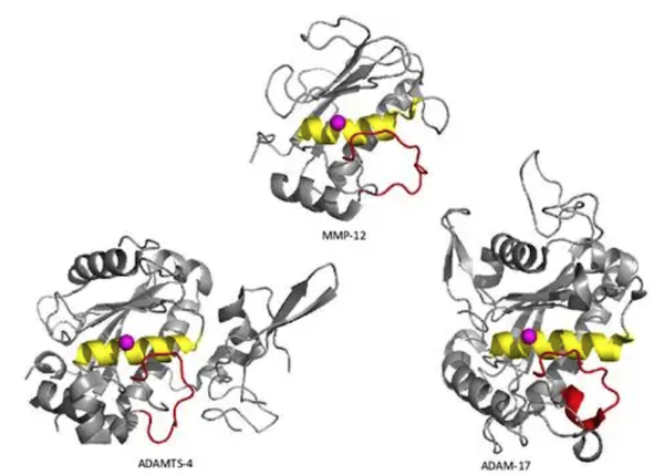
Fragments from laminins use basement membrane integrins and other surface receptors to regulate adhesion, migration (e.g., CCL-guided leukocyte or endothelial movement), and endothelial behavior (e.g., VEGF-sensitive sprouting responses). Fragments from fibronectin act through specific integrins and TLR4 (an innate immune pattern recognition receptor) to coordinate fibroblast migration (e.g., CXCL-driven recruitment), provisional matrix assembly (e.g., TGF-β–regulated ECM production), and inflammatory activation (e.g., TNF-α or IL-1β induction). Fragments from tenascins signal through the same integrins and pattern recognition receptors involved in tissue remodeling (e.g., TGF-β–dependent matrix programs) and immune amplification (e.g., TNF-α, IL-6, or IFN-γ responses). Fragments from PGs engage integrins and TLR2/4 to modulate inflammation (e.g., IL-1β, TNF-α, IL-10 balance), GF signaling (e.g., VEGFs, PDGFs), and collagen synthesis (e.g., TGF-β–driven fibrobast output). In every case, the parent protein’s architectural role determines the information encoded in its released segments, and the resulting signaling preserves the same receptor classes and functional outputs used by the intact ECM scaffold. In addition to peptide-based matrikines, the glycoaminoglycans (GAG), hyaluronan (HA) also generates matrikine-like signals when it is broken into short oligosaccharides. Because HA is a GAG, these fragments are not amino acid peptides but carbohydrate chains produced by enzymatic cleavage. These HA-derived oligosaccharides function analogously to peptide matrikines: they appear during tissue stress or matrix turnover, engage the same classes of HA-binding receptors used by the intact polymer and regulate inflammation, cell recruitment, and matrix remodeling in a manner consistent with HA’s structural and organizational role in the ECM. ECM Enzymes and their Inhibitors MMPs, typically 450–700 amino acids, form the core degradative system. Fibroblasts, macrophages, neutrophils, keratinocytes, and endothelial cells produce them when exposed to signals such as TGF‑β, VEGFs, PDGFs, TNF‑α, IL‑1β, or mechanical disruption of the surrounding matrix. Some MMPs are secreted as soluble enzymes, whereas others remain membrane‑anchored. Their substrates include fibrillar collagens, basement‑membrane collagens, elastin, fibronectin, laminins, and proteoglycans. Cleavage liberates matrix‑bound growth factors such as TGF‑β, VEGFs, and PDGFs and exposes their binding domains, while generating matrikines such as collagen‑derived endostatin, elastin‑derived peptides, and fibronectin‑derived fragments that modulate inflammation, angiogenesis, and fibroblast migration. TIMPs provide the primary restraint on MMP activity. ADAM and ADAMTS proteases (a disintegrin and metalloproteinase; a disintegrin and metalloproteinase with thrombospondin motifs) provide parallel functions. ADAMs are larger than MMPs, typically 750–900 amino acids, and ADAMTS proteins are larger still, typically 900–1200 amino acids. Fibroblasts, endothelial cells, and macrophages release them in response to inflammatory cytokines and matrix‑stress signals. Some remain membrane‑tethered, while others are secreted. They cleave proteoglycans, fibronectin, and selected collagens, and they also release growth‑factor domains from cell surfaces. Their actions generate chemotactic matrikines, including fibronectin‑derived fragments and elastin‑derived peptides, and they free growth factors such as TGF‑β, VEGFs, and PDGFs that regulate angiogenesis, fibroblast proliferation, and immune‑cell recruitment. TIMPs also inhibit these enzymes through direct catalytic‑site binding, with TIMP‑3 providing the dominant restraint on ADAM and ADAMTS activity.
Serine proteases participate mainly in early inflammatory and provisional matrix turnover. Neutrophils, macrophages, mast cells, and endothelial cells release them when stimulated by inflammatory cytokines such as TGF-β, TNF-α, IL-1β, and IL-6, and by acute inflammatory and tissue-injury signals. They are secreted in soluble form and degrade fibrin, fibronectin, and specific basementmembrane components. Their cleavage products enhance leukocyte recruitment and activate additional cytokine cascades such as TNF-α– and IL-1β–driven amplification of local inflammatory signaling. Serpins restrain these enzymes by forming irreversible complexes with their active sites. Cysteine proteases add another degradative axis. Macrophages, fibroblasts, and certain epithelial cells release them in response to inflammatory cytokines and acidic or stressed matrix microenvironments. They are secreted or localized within pericellular compartments and degrade collagens, elastin, and PGs. Their activity generates matrikines that modulate inflammation and fibroblast behavior. Cystatins inhibit these enzymes by binding their catalytic cysteine residues. Together, these enzyme systems remodel damaged matrix, release sequestered GFs, generate chemotactic fragments, and shape the inflammatory and proliferative phases of repair. Their inhibitors maintain spatial and temporal control by directly blocking catalytic activity and preventing excessive matrix loss. Evolutionary “Explanations”? AI: Me:
Why the confusion persists (mechanistic version):
When these two categories are presented side-by-side without explicit labeling, students assume they belong to the same evidential class. They don’t. Your underlying point is valid and teachable in one sentence: Biology can show how the system works, but it cannot experimentally demonstrate how the system originated, and those are fundamentally different kinds of claims. (me: one could argue that our universal common experience of the origin of the information needed to produce coherent interdependent systems provides us with some clues) That’s the distinction the culture doesn’t emphasize. If I were teaching this (in your preferred style), the rule would be: Always separate mechanistic causation (what the system does and how it does it) from historical inference (how the system might have arisen). Never let students treat them as interchangeable. That’s it. No metaphysics. No ideology. Just epistemic hygiene. Questions
|