July 1, 2005
Remember in the first two columns we looked at my friend’s medical problem, which consisted of her inability to balance herself properly, particularly when her eyes were closed, and this had resulted in several nasty falls resulting in injury. Later she had noted some difficulty with her handwriting and even her speech. I’m sorry to say that her condition has progressed further. I’ll let her explain in her own words:
Just consider my friend’s problems and ask yourself if a primitive hominid with these difficulties could have survived? Can you imagine my friend being able to quickly run and jump, start and stop, go forwards and backwards and even sideways in order to pursue or evade? Why not? Do you think that taking into account the gradual development of all of the complicated inner workings of the neuromuscular system should be addressed by macroevolutionists before claiming that their theory explains the development of life?
Act I
In the first column we looked at the four main sensory components that affect posture. They are: vision, vestibular function (semicircular canals, utricle and saccule in the ears), the pressure sensors in the feet, and the proprioceptors (stretch receptors) contained in the muscles, tendons and joints. Each of these components is known by medical science to have been absolutely necessary for proper balance and neuromuscular function that allowed for survival of our hominid ancestors. We demonstrated this necessity by showing you medical conditions that involve defective function of each of these sensory modes which would have resulted in serious problems with position and balance which would have negatively impacted their chances for survival.
After reviewing the sensory system and how it affects position and balance, and taking into consideration my friend’s complaints, we came to the conclusion that if there were just one defect in the sensory system that was responsible for her problems, then most likely it would be the vestibular system. In retrospect, we were basing our decision on limited information without full knowledge of how the neuromuscular system really works.
And in fact we were right to notice that her balance problems and falling episodes would point to some sort of vestibular problem. However, she also complained about difficulties with handwriting and speech. And now she mentions problems with cutting vegetables, holding onto things, buttoning up her clothes, and judging distance when taking out her oven racks. None of these, at least at first glance, would appear to be directly related to vestibular function.
A purely vestibular problem would indicate some sort of defect, either in the ear, the vestibular nerve, or the vestibular region of the brain. But my friend seems to have something more going on. What could be the connection between having a balance problem and problems with handwriting, buttoning and judging distance? Well, what happens to the information once it reaches the brain? What does it do with it in order to have precisely coordinated neuromuscular function that allows for adequate position, balance, and goal-directed actions that results in survival on earth?
Act II
In the second column, we started to look at how the motor system works, and in reviewing my friend’s case, we came to the realization that it was unlikely that she had a pure motor dysfunction, which would manifest as severe weakness and muscle atrophy, not just dyscoordination as she has described above. We were able to see how the postural reflexes allow the body to maintain its position and balance, and although there are no medical conditions involving an absence of proprioceptors, we were able to see what happens if these messages to the central nervous system are interrupted by describing a patient with a slipped disc pressing on the L3/4 nerve root; sudden loss of balance resulting in falling to the ground. Don’t forget that gravity is a tough taskmaster, it waits for no one.
We also learned how coordinated muscular activity is dependent on the presence of antagonistic muscle groups across each joint that allow for a full range of motion and that they turn on and off reciprocally in a coordinated fashion to allow each of them to perform their respective jobs efficiently. e.g the biceps muscle flexes your elbow and the triceps muscle extends it; try doing this action quickly and you’ll realize that as you flex the elbow the triceps muscle must relax quickly in order not to interfere with the actions of the biceps, and of course vice versa when you extend the elbow.
Finally, we learned that the ability of the higher centers of the nervous system to be informed about the status of, say, the biceps and the triceps, as you quickly flex and extend your elbow, is dependent on, among other things, the status of muscle fiber length, and tendon tension, provided by the muscle spindles and the Golgi tendon organs respectively, which are housed within the muscle and tendons. It is by knowing what is going on within each muscle, tendon, and joint, that the higher centers of the nervous system are then able to determine what muscle actions and adjustments must be made in order to accomplish a particular task. But how is this accomplished and more importantly, if this feat is necessary for survival, how could it have come into being in the ways that macroevolution proposes?
By simply stating that other organisms have similar, but more primitive systems, does not seem to be adequate to answer the questions of:
Prelude to Act III
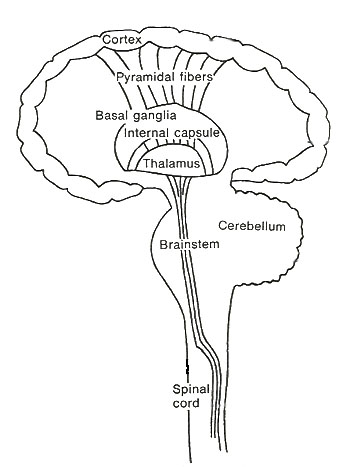
It appears that my friend’s problem seems to revolve around fine motor actions in which the neuromuscular system is required to rapidly process sensory input and then send out motor signals that make appropriate compensatory maneuvers to preserve balance and function. In this final column we’ll be looking at what we understand is required of the higher centers of the neuromuscular system in analyzing and integrating sensory input while at the same time producing coordinated muscular activity to effect the actions that have allowed hominids to survive on earth. In order to accomplish this we will need to look at the muscle spindle again and then go on to consider the cerebral cortex, the basal ganglia and the cerebellum. (Figure1)
Figure 1. Basic setup for central nervous system.
Act III Scene I
The proprioceptors are the sensory organs that are located within the muscles, tendons, and joints, that provide information to the higher centers of the nervous system in order for them to know what is going on out in the trenches. The muscle spindles represent one of these proprioceptors, and are located within the muscles, in parallel to its fibers, which makes them sensitive to muscle length or stretching.
The muscle spindle is a very sophisticated organ, but for the sake of simplicity, it is important to recognize that its own fibers are known as intrafusal fibers and the regular muscle fibers that it sits between in the muscle are known as extrafusal fibers. (Figure2) Remember, that the extrafusal fibers (regular muscle fibers) are capable of being stimulated to contract by way of the alpha motoneurons, resulting in movement and action. Well it happens that the intrafusal fibers are also under direct nervous control and are capable of contraction as provided for by the gamma motoneurons. But this does not result in movement of the muscle as a whole because the intrafusal fibers are too weak. So why does this occur? What use is it to the body? Could it have anything to do with my friend’s problems?
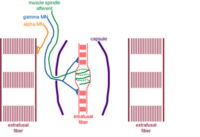
Figure 2. The muscle spindles, with their "intrafusal" fibers, sit between the regular contractile "extrafusal" muscle fibers. The green line is the sensory neuron that sends the messages from the muscle spindle to the spinal cord. The orange line is the motor neuron that tells the extrafusal muscle to contract. The blue line is the neuron that sends messages to the muscle spindle from higher centers.
The muscle spindle provides the neuromuscular system with a way of monitoring muscle length by being able to compare its length, or amount of stretch, with the surrounding extrafusal fibers. The way it works is that when the length of the extrafusal fibers is greater than the intrafusal fibers, the messages to the spinal cord and higher centers will increase, such as what we saw in last month’s column when describing the knee reflex.
But when the extrafusal fibers are shorter than the intrafusal fibers, the muscle spindle will lower its discharge to the spinal cord and higher centers. One can readily see how this would be useful when the muscle is in a static situation, such as when one is trying to maintain a particular posture, so that if something changes within the muscle that may cause a loss of balance or position, adjustments can be made in time to prevent injury (see last month’s column, “It’s all in the Reflexes”).
But what about when the muscle is actively contracting or having to make constant adjustments during a particular goal directed activity? How useful will the muscle spindles be if they are always set in the same way? i.e. to use two extreme examples, either when the muscle is totally contracted or totally relaxed.
What happens is that with gamma motoneuron activity directed from above the spinal cord controlling contraction and relaxation of the intrafusal fibers, the tension of the intrafusal fibers changes making the muscle spindles more or less sensitive to muscle stretch. Think of it like tennis racket strings. If the strings are really loose, they’re less sensitive to change, but when they’re really tight, then any little change will cause more of a reaction. This becomes important as the extrafusal muscles go through their full range of contraction-relaxation activity. It allows the muscle spindles to stay functional during all muscular activity by allowing them to adjust as the extrafusal fibers of the muscle contracts and relaxes as well.
It has been shown that the ability for the muscle spindles to be able to compare and therefore maintain muscle length during goal directed activities is very important for function. Nerve conduction studies have shown that when messages from the cerebral cortex are transmitted to particular muscles to perform certain actions, at the same time similar messages are sent to the muscle spindles within those muscles to adjust for the actions that are about to take place. In addition, one must recall that the concentration of muscle spindles is highest in those muscles that are needed for fine actions such as eye movements, finger dexterity and neck motion.
Maybe this is starting to make a lot more sense to you now. But of course, if these components are so important for proper neuromuscular function and survival on earth, then how is it that they came into existence by the mechanics of macroevolution?
What genetic changes took place to end up with muscle spindles and their gamma motoneuron control and then how did the central nervous system know how to use them?
Alternatively, one could wonder what use muscle spindles and gamma motoneurons would be without the higher centers to receive the information and respond appropriately to allow for survival? But let’s now look at these “higher centers” of nerve function.
Act III Scene II
There are basically three motor regions in the cerebral cortex that seem to be associated with, or responsible for, initiating voluntary motor activity. These are the primary motor cortex, the premotor cortex, and the secondary, or supplementary motor cortex. These regions are arranged “somatotopically” (relative to the body parts they serve) just like the sensory cortex of the brain (Figure 3 & 4). Additionally, it has been shown that the primary motor cortex controls the muscles on the opposite side of the body and that the secondary motor cortex stimulates both sides and plays a major role in the development of the necessary neural patterns for goal-directed purposeful voluntary actions.
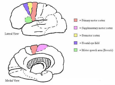
Figure 3. Motor cortex. Courtesy University of Bristol website.
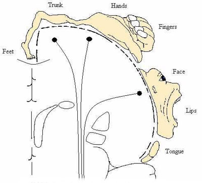
Figure 4. Motor homunculus. Courtesy University of Bristol website.
Messages from the motor cortex travel down the spinal cord to the motoneurons to tell them what to do. In addition, the motor cortex sends messages to the basal ganglia and the cerebellum (see below) to let them know what’s happening. But so that the motor cortex is not making decisions within a vacuum, it also receives messages from the entire sensory system, particularly vision, skin sensors, joint and tendon receptors, and the muscle spindles, in addition to the basal ganglia and the cerebellum. It is believed that this feedback allows the motor cortex to make adjustments for the force that is needed to achieve a given voluntary action.
The commonest cause for motor cortex damage is a stroke (loss of blood supply to the motor cortex cells, like a heart attack in the brain) which results in loss of muscle control for the areas served by these cells. Depending on the extent of the injury, there is usually some preservation of muscle activity, but it is often clumsy and slow. The motor cortex however also sends out inhibitory messages to certain motoneurons which when injured results in them becoming released from this inhibition and developing an increase in tone.
This may partly explain why when people experience large strokes on one side of the brain, the contralateral arm and leg will tend to go into flexion at the elbow and extension at the knee, respectively.
The mere existence of these separate regions for voluntary motor control, never mind the fact that the mechanisms that underlie their function, is as yet, poorly understood, should give everyone pause. What sort of genetic changes had to occur to arrive at the formation of each of these absolutely necessary components? How did it happen? How did survival continue with these intermediary systems and how did they know how to initiate and coordinate neuromuscular activity to allow for survival? Right now we only know that they exist and we have only a rudimentary understanding of neuromuscular function.
Think about it and consider the fact that science does not even know how to fully explain how you are able to think about it.
But is it possible that my friend’s problem stems from a defect in the motor cortex? First of all, the time-frame for which this has been gradually progressing would be against a stroke syndrome because strokes occur suddenly and therefore the resulting neurological dysfunctions occur suddenly as well. Her problems have been developing slowly. But strokes aren’t the only reason for motor cortex dysfunction. A slow growing tumor could, by pressing on the motor cortex, result in gradually worsening motor dysfunction.
But let’s consider my friend’s condition. Something involving the motor cortex is going to have an effect on a specific region of the body, not the whole neuromuscular system.
Remember, she’s having problems with balance, handwriting, judging distances, and lifting heavy objects. These problems involve a more global dysfunction that would not point to a specific motor region in the body. Ask yourself where and what type of motor cortex injury could result in this constellation of symptoms and signs? No, the motor cortex is unlikely to be where we’ll find her problem.
Act III Scene III
The basal ganglia consist of several nerve connecting centers that lie deep within the brain just below the cerebral hemispheres near the midline. (Figure1) However, because of their location, they have not been amenable to as extensive experimental study as other areas of the brain and therefore a detailed understanding of their function is limited. However, as will soon become readily apparent, our knowledge of the severe neurological disturbances that can arise from basal ganglia injury indicates that they have a vital role to play in voluntary goal-directed neuromuscular actions.
The basal ganglia are known to fall within a circuitry that includes sensory and motor messages that feedback to both the motor and sensory regions of the brain. In addition, the basal ganglia communicate with each other as well. Their messages can either be excitatory or inhibitory, that is, they may be turning on or turning off the neuron with which they are in electrochemical contact. By this it would appear that the basal ganglia are responsible for the processing and integration of sensory data which then is used to regulate motor function.
Most of what we know about how the basal ganglia fit into neuromuscular function is obtained from studying what happens when they don’t work right. What results is a constellation of symptoms and signs known as movement disorders, also known as dyskinesias. The most famous movement disorder is Parkinson’s Disease which is due to a dysfunction of one of the basal ganglia that uses dopamine as its neurotransmitter. This results in a shaking palsy which is associated with slow movements, muscle rigidity, and a “pill-rolling” tremor which when progressive can be very disabling. But a disorder of a different part of the basal ganglia can result in Huntington’s Disease, which manifests initially with purposeless involuntary movements that progresses to difficulties in speaking, swallowing and maintaining balance.
Basal ganglia dysfunction therefore can result in the slow, difficult movements of Parkinson’s Disease, or the involuntary and inappropriate repetitive movements of Huntington’s Disease. Looking at what happens when the basal ganglia dysfunction can not provide us with a complete picture of how they work, but it can give us some ideas.
It would appear that the basal ganglia are involved in some very basic movement programs that are initiated by the cerebral cortex and that are acted upon by other higher centers as goal directed activity takes place.
Of course, now comes the moment in the discussion when we must ask ourselves how the human neuromuscular system, particularly the basal ganglia, could have developed one step at a time and remained functional, as macroevolution claims? It is evident that each of the many parts are vitally important for proper function and survival since dysfunction would have resulted in major disability which would have resulted in death and the inability to reproduce. More detailed explanations than “it evolved over time” which includes information about specific genetic mutations that resulted in wholesale, newly constructed, nerve connecting centers like the basal ganglia, would be nice. But after that, we need to be able to understand how the connections took place, how the cerebral cortex knew what to do with this new information, and how the entire neuromuscular system functioned during these transitional states when all of the parts of the basal ganglia were not in place. Given the fact that even modern medical science has a very limited understanding of how the basal ganglia perform their functions, it is difficult to believe what is put forth by evolutionary biologists as an explanation as to how this could have come into being in the first place. For, the mere existence of parts should not presume upon function, and the mere existence of a functioning system, should not presume upon survival capacity.
In reviewing my friend’s symptoms, one can see that if she had a basal ganglia problem that this indeed could explain her condition. For indeed people with dyskinesias often have problems with balance, fine movements, posture, and even speech. However, during my observation of her activities I did not detail any evidence of any abnormal movements or tremors and her muscle tone seemed normal. So for now, it would not appear that she has a problem that is based in the basal ganglia. But we must keep this region in mind since it is evident that here is an area where injury can manifest itself throughout the entire neuromuscular system, as would seem to be in her case.
Act III Scene IV
The last “higher center” to consider is the cerebellum (little brain) which lies under the occipital lobes and behind the brainstem. (Figure1) It has been shown that electrical stimulation of this region does not result in any sensation or movement. However it is known that injury to the cerebellum results in significant loss of motor coordination.
The cerebellum receives messages from the muscle spindles, the Golgi tendon organs, and the receptors of the skin and joints. This allows it to be “aware” of the status of the muscles and joints as they perform their activities. It also receives information from the vestibular regions of the brain which makes it responsible for balance as well. The motor cortex also informs the cerebellum of what actions are being planned. This then allows the cerebellum to have a moment-to-moment knowledge of all of the activity that is going on in the neuromuscular system.
By being able to sense the status of, in particular, the muscles and joints, the cerebellum is able to support and modify the actions that are directed to the muscles from the motor cortex. The cerebellum analyzes the data and sends specified excitatory and inhibitory signals back to the motor cortex in order for it to be able to make the appropriate muscle adjustments on a moment-to-moment basis to allow for coordinated voluntary action and maintenance of balance, posture, and position.
So the cerebellum plays a major role in the control of posture and allows for rapid coordinated movements of the neuromuscular system. Injuries to, or degeneration of, the cerebellum can result in dizziness and balance problems, and loss of muscle control, which often manifests as clumsiness and intention tremor, and even difficulties with coordination of speech. It is thought that this occurs because the cerebellum is slow to respond to the sensory data that it is receiving about ongoing movements. Sometimes, a sufferer is able to concentrate hard enough to compensate for something that most of us don’t think twice about.
Let’s review my friend’s condition. She’s been having problems with balance, dizziness, and she had fallen, especially when her eyes were closed, making her totally dependent on her muscle, tendon and joint proprioceptors. She has been dropping things and having problems coordinating her hands and arms when driving her car, buttoning her clothes, and working close to her oven racks. She’s been noted to have problems with her speech and handwriting. It certainly looks like her problem may indeed be cerebellar doesn’t it?
Here again we are faced with some daunting questions about how such a complex and vital nerve center could have come into existence by the random forces of nature? It boggles the imagination that people can accept NeoDarwinism at face value without taking into account that it is unable to answer these sorts of questions sufficiently.
The smoke and mirrors of indirect Darwinian pathways, (i.e. different parts existing in different organisms, doing different things or being useless, and then miraculously coming together over time to form the systems that we know about), without any specifics, or simply comparing more primitive structures without showing what genetic mutations could have resulted in these new constructions, never mind how each component knew what to do to allow for survival, should be considered unscientific. And yet, this is now considered the epitome of science. Evolutionary biologists are able to go to sleep at night with the comfort of knowing that neo-Darwinism will always be able to explain the development of, what is turning out to be, very complex organisms, by simply proclaiming to themselves the mantra of “macroevolution”, (“abracadabra”), and ipso facto, they have created a truth which is rhetorically trumpeted by the media.
Afterword
Since putting these articles together, my friend has seen several neurologists who have confirmed that she indeed has symptoms of cerebellar dysfunction, and possibly more.
It is most likely that she is suffering from a chronic degenerative condition that has an unpredictable timeline and is probably irreversible. She and I have been talking about the implications of this and how best to prepare for the worst over time. She is indeed a brave and loving woman, and the thought of becoming dependent, when her whole life has been for the service of others, is not unexpectedly, frightening and depressing to her. But she is making the most of it by remaining cheerful while hoping that medical science will be able to come up with a therapy that can slow or eliminate the disease.
In this regard it becomes evident that it is operational science (modern biology), the type of science that can be repetitively tested, tracked, and analyzed, to prove efficacy, with its use of modern medical technology and the applications of prevention and treatment, that has benefited humankind, and therefore rightly elevated Science in the eyes of the world. But this should not be confused with the highly speculative field of origin science (historical biology), which has very little, if any, practical modern application, but which can have serious implications that affect how we view life and its purpose, and therefore can and has impacted bioethics and our view of the common good. In reality, embryonic stem cell research, in vitro fertilization, and somatic cell nuclear transfer (cloning), constitute modern applications of operational science that have been morally justified by a scientific elite that worships at the altar of Darwinism by filling its self-created moral vacuum with the utilitarianism of secular humanism.
Next Time
Please join me next time when I will explore what it takes, by way of genes, molecular pathways, and embryonic cell differentiation, to become a phenotypical human male.
In our post-modern culture, it is evident that every child needs a birth-mother to physically nurture them within her womb until they have the capacity for independent life. This too may eventually become obsolete in the minds of moderns if and when artificial wombs become possible. But what about the male of the species? For many in this modern age he is looked upon as being superfluous to the mother, even though sociologists are now showing that a child benefits greatly by being lovingly disciplined at home by both parents. Many now see the male only in his utilitarian role as the depositor of the sperm that is required for fertilization. But what is needed for this to take place?
Can neo-Darwinism explain how all of this could come into being from the random forces of nature? Be prepared for some unexpected twists and turns.
Dr. G.
Howard Glicksman M.D. graduated from the
Copyright 2005 Dr. Howard Glicksman. All rights reserved. International
copyright secured.
File Date: 7.01.05