December 1, 2004
Vision is a complex process. Two months ago we reviewed how the eye is able to allow light to enter it and be focused on the retina. Then last month we detailed how the retina is able to generate nerve impulses that travel to the brain for the interpretation of “sight”.
This month we look at how these visual messages are distributed and organized within the brain in order to create a particular neuroexcitatory spatial pattern for analysis.
The brain is the central processing unit that interprets all of the neurological messages that come in from all over the body. The eye represents a peripheral unit, like any other sensory organ in the body. It’s out in the trenches doing reconnaissance for the brain. Central blindness refers to when the eyes work fine but it’s the brain that isn’t doing the job of processing the visual information correctly.
The Path Revealed
Each optic nerve consists of about one million axons that have come from the ganglion cells. Now don’t forget that the ganglion cells are simply transmitting the messages that they received from the bipolar cells and they in turn from the rods and cones. It’s sort of like a huge neurobiomolecular relay race. The final goal is to reach the visual center of the brain where some sort of spatial pattern of nerve excitation representing the original light source that started the race is finally processed and interpreted as “vision.”
About 80% of the axons from the ganglion cells in the optic nerve travel to a junction box in the brain called the lateral geniculate body. In this nerve connecting center each ganglion axon passes on its message by way of the release of a neurotransmitter that stimulates another neuron that now takes that message to the visual cortex further back in the brain.
The other 20% of the ganglion axons veer off just before the junction box and join up with another system which is responsible for some of the automatic reflexes that occur in the eye. When light is shone into the eye this causes the pupil reflexively to become smaller, and when there is less light, as in a dark room, the pupil automatically opens up to allow more light in. It is the messages from these ganglion cells that start the reflex arc that results in these actions.
Who’s on First !?
Having said all this, we now need to look at the question of which ganglion axons, carrying messages from the retina, go where? It would seem to make sense that all of the messages from one eye would end up in one visual cortex and all of the ones from the other would end up in the other, wouldn’t it? Otherwise, how can the brain interpret all of the messages if they’re mixed up? In order to understand what actually happens and how this affects our sight we must first comment on how lenses affect the images that they refract and also provide you with a frame of reference for the discussion of vision.
Reversal of Reality: Focusing Side-effects
To begin with, we must consider the nature of the image that is projected on the retina after the light rays travel through the eye. If you’ve ever played around with lenses you may remember that each time light rays go through a curved surface, the rays not only are bent but the image on the other side becomes totally reversed.
Therefore, when we consider what happens to the image of light as it goes through the eye, we must take into account the fact that the light undergoes three separate refractions. The first refraction occurs as the light travels through the cornea. At this point, the image would be totally reversed, meaning that it would be turned around and upside down. But don’t forget, the light still needs to go through the lens before it travels on to the retina.
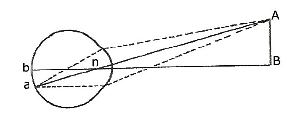
The lens has two convex surfaces as opposed to the cornea’s one. The image travels through the front surface of the lens and in doing so would be set right again. But then it is further refracted as it travels through the back surface of the lens, which results in the final retinal image being reversed; that is, turned around and upside down. (See Figure 1)
Figure 1. AB in visual field is reversed and upside down on retina.
You may be wondering how this could affect our vision? Don’t forget, that the retinal photoreceptor cells simply send an image to the brain that is based on the light that reflects off of the object that is being focused on. Therefore, if the image itself has been reversed, that is turned around and upside down, the message that is sent from the retina to the brain will reflect this as well. It’s up to the brain to be able to decipher this mirror-image electrical message that’s being sent from the eyes.
Frames of Reference
In order to understand the discussion going forward it’s important for you to understand some terms of reference regarding the visual fields and the regions of the retina. The visual fields of each eye, (that which is being observed), can be divided vertically into right and left fields. In a similar fashion, the retina of each eye, (that which does the observing), can also be divided into a right and left region by drawing an imaginary line from the top to bottom of the eye through the fovea. (Additionally, each field and region of the retina can also be divided into a upper (superior) and lower (inferior) half )
But because each eye is already designated as “right” and “left,” it would be confusing for scientists to refer to the visual fields or each region of the retina of either eye by “right” and “left” as well. So, we need a better way of clearly differentiating between the visual field being observed, the part of the retina that is doing the observing, and in which eye all of this is taking place.
The temporal bone is the outside boundary of each eye; that is, it is to the left of the left eye and to the right of the right eye. Similarly, the nose is the inside boundary for each eye; that is, it is to the right of the left eye and to the left of the right eye. Therefore, each vertical half-field of vision is called either the temporal or the nasalfield.
The temporal visual field in the left eye is the far left half-field and the temporal half-field in the right eye is the far right half-field. Similarly, the nasal visual field of the left eye is the inside or right half-field and the nasal field of the right eye is the inside or left half-field. (See Figure 2)
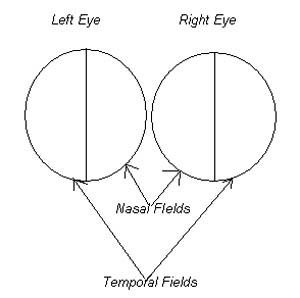
Figure 2. Visual fields.
In a similar fashion, when we discuss the retina we are talking about where the retina actually sits in the eye. Therefore, the temporal retina of the left eye sits in the outside or far left-half of the back of the eyeball and the nasal retina of the left eye sits in the inside or right-half of the retinal field in the left eye. Similarly, the temporal retina of the right eye sits in the outside or far right-half of the back of the eyeball and the nasal retina of the right eye sits in the inside or left-half of the retinal field in the right eye.
What’s on Second !?
When we consider the relationship between what is seen within the visual field of a particular eye and where its image is on that eye’s retina, we must keep in mind that the image will be reversed and turned upside down. Therefore, whatever is in the temporal field of vision of either eye will always be imaged on the nasal retina and whatever is in the nasal field of vision in either eye will always be imaged on the temporal retina. (Additionally, for those of you who are ahead of the others: whatever is seen in the superior field will be imaged on the inferior retina and whatever is seen in the inferior field will be imaged on the superior retina)
It’s All a Matter of Perspective
One more important thing to remember about vision can be demonstrated with this exercise. If you focus on an object and then alternately look at it with each eye, you’ll notice that there is a significant overlap between each eye’s nasal fields, from a slightly different angle. This means that when you focus on something, the eye is capable of sending messages to the brain that give it two different perspectives. This is how we are able to achieve our depth perception.
Split Screen: Crisscrossing the Neurobiomolecular Highway
Now that you understand this aspect of our vision we can go on to discuss where the messages go in the brain. In reality if you imagine a vertical line going through the fovea, all of the photoreceptors to the right in both eyes; that is, the nasal retina in the left eye and the temporal retina in the right eye: send their messages to ganglion cells that send their axons to the right side of the brain.
Similarly, all of the photoreceptor cells to the left of the fovea in both eyes; that is, the temporal retina in the left eye and the nasal retina in the right eye: send their messages to ganglion cells that send their axons to the left side of the brain.
In order for this to happen, all of the messages from the temporal retina of both the right and left eyes stay on the right and left side of the brain respectively. While all of the messages from the nasal retina of both the right and left eyes must cross over to the left and right sides of the brain respectively. This all crosses at what is called the optic chiasm. (See Figure 3)
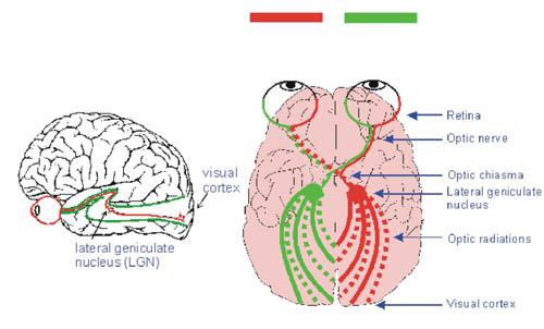
Figure 3. Obtained with permission from www.yorku.ca website
If you remember which half of the retina “sees” which visual field, you’ll come to realize that everything in the left-half of the visual field of both eyes goes to the right side of the brain and everything in the right-half of the visual field of both eyes goes to the left side of the brain. Remember, I said that because the image that goes through the eye is reversed by the combined effects of the cornea and the lens, everything in the temporal field will be imaged on the nasal retina and everything in the nasal field will be imaged on the temporal retina.
This means that something in the temporal, or left half-field of the left eye, will image on the nasal or right-half of the left retina. But we just said that all of the messages from the nasal retina cross from the left side to the right side of the brain. So the visual messages that are a result of the left-half field of left eye vision stimulating its nasal retina will be sent to the right visual cortex of the brain. Left-half vision goes to the right brain.
Similarly, something in the temporal, or right half-field of the right eye, will image on the nasal, or left-half of the right retina. But once again, we know that all of the messages from the nasal retina of the right eye cross over to the left side of the brain. So in this case the visual messages that are a result of the right half-field of right eye vision stimulating its nasal retina will be sent to the left visual cortex of the brain. Right-half vision goes to the left brain. (See Figure 3)
If we look at the nasal fields we see the same thing happens. Something in the nasal, or right-hand field of the left eye, will image on the temporal, or left-half of the left retina. However, we know that all messages from the temporal retina stay on their same side.
Therefore in this case, the messages from the left temporal retina will be sent to the left brain. Once again, right-half vision goes to the left brain.
Finally, something in the nasal, or left-hand field of the right eye, will image on the temporal, or right-half of the right retina. Images from the temporal retina stay on the same side, so these would be sent to the right brain. Hence, left-half vision ends up in the right brain again.
I Don’t Know’s on Third!
The brain then takes this, turned around, upside down, split-up, and overlapping con-glomeration of photon-generated impulses, that have originated in the retina, through the bipolar cells, the ganglion cells, the lateral geniculate body and on to the occipital lobes, and produces what we call “vision”. Here’s how it does it.
????????????????????????????????????????????????????????????????????????
????????????????????????????????????????????????????????????????????????
????????????????????????????????????????????????????????????????????????
????????????????????????????????????????????????????????????????????????
????????????????????????????????????????????????????????????????????????
No one really understands exactly how we are able to see. It’s sort of like asking ourselves what is the neurobiomolecular basis for a given thought, desire, or emotion.
We may be able to ascertain where in the brain these processes may be taking place, and even which neurotransmitters, in what concentrations, and with what other neurons, reactions are occurring. But we still do not understand exactly how these processes result in special sensations like vision much less a given thought.
We just don’t understand how it is that we can understand. The philosopher Gabriel Marcel defined a mystery as: “a problem that encroaches on its own data”. By that he meant that the questioner unwittingly becomes the object of the question. The human brain is trying to figure out how itself works.
Evolutionary Simplicity?
Review of this and the last two columns clearly demonstrates:
the extreme complexity and physiological interdependence of many parts of the eyeball
the absolute necessity of many specific biomolecules reacting in exactly the right order to allow for photoreceptor cells and other neurons to transmit nervous impulses to the brain
the presence of, not only an eyeball whose size is in the proper order to allow for focusing by the cornea and lens, but also a region in the retina (fovea) that is outfitted with the proper concentration of photoreceptor cells that are connected to the brain in a 1:1:1 fashion to allow for clear vision
that vision is dependent on a complex array of turned around, upside down, split-up, and overlapping messages, from over two million optic nerve fibers that course their way to the visual cortex causing a neuroexcitatory spatial pattern that is interpreted as sight
that scientists are blind to how the brain accomplishes the task of vision
The foregoing is likely to give most people pause before they subscribe to the theory of macroevolution and how it may apply to the development of the human eye and vision. How can one be so certain of an origins theory when one still doesn’t fully understand how something actually works? Most of what I’ve read by its supporters about this topic contains a lot of rhetoric and assumption without much detail and logical progression. It all seems just a bit premature and somewhat presumptuous.
Quite frankly, science just doesn’t have the tools to be able to definitively make this determination, yet. Will it ever have them? Maybe yes, maybe no. Until such time, I reserve the right to look upon evolutionary biologists’ explanations for the development of the human eye and the sensation of vision, with a large amount of skepticism, and as seeming overly simplistic and in need of a heavy dose of blind faith.
Next month we’ll be looking at the ear and hearing. It should provide us with more things to wonder about and more questions to think about for macroevolution.
Join me then in: Wired for Much More than Sound: Part VII—The Ear and Hearing
Dr. G.
Howard Glicksman M. D. graduated from the University of Toronto in 1978. He practiced primary care medicine for almost 25 yrs in Oakville, Ontario and Spring Hill, Florida. He recently left his private practice and has started to practice palliative medicine for a Hospice organization in his community. He has a special interest in how the ethos of our culture has been influenced by modern science’s understanding and promotion of what it means to be a human being.
Copyright 2004 Dr. Howard Glicksman. All rights reserved. International
copyright secured.
File Date:12.01.04