July 1, 2004
In the time that it takes for you to read this paragraph; your automatic pacemaker has stimulated your heart to contract several times; a message from your brainstem has traveled through various nerves to stimulate your diaphragm to make you breathe at least once; and the nerves that control your eye muscles have allowed you to scan and focus on what you have just read. Pretty amazing isn't it?
But there's much more going on than that. For if you are like most people, you're probably sitting at your computer right now, trying to decide whether to read the whole column now or print it off. In order to be able to use your mouse or keyboard to scroll down, your brain would have to be sending the proper messages to the right muscles. And, for you to be able to sit erect in your chair, your spinal muscles would have to be working in a coordinated fashion, orchestrated by the brain. And finally, as you move your head up and down and left or right, as you search for your mouse and decide to turn on your copier, you wouldn't be able to keep your position in space if it weren't for the properly working balance organs in your ears, under the control of the brain.
How do we know all of this? Well, if your automatic pacemaker doesn't work, you usually develop a very slow heart rate generated by the ventricle and you often pass out. That's why medical science implants pacemakers. If a person suffers an major injury to the brainstem or upper neck, this usually damages the respiratory center or nerves for respiration, and the person dies of asphyxia. Diabetics sometimes develop weakness of one or more of the cranial nerves that are responsible for movements of the eye. When this happens, the person is not able to move the eyes in a coordinated fashion, and they see double and can't focus three dimensionally..
If a person suffers an injury to the dominant lobe of the brain, they wouldn't be able to use their dominant hand for moving the mouse or scrolling down. If a person suffers an injury to a particular part of the cerebellum, they may have great difficulty being able to stay in a sitting position without support. Finally, if a person suffers from dysfunction of the balance organ in the ear, they may find that with any minimal head movement they will suffer from severe vertigo and vomiting.
So how is the body capable of performing all of these feats? Most people would say that it's the brain, the nerves and muscles of the body that are responsible for all of these actions. And they would be right. But that would only be answering the what and not the how. Let me explain further. The next two months' columns will deal with nerve cells. This month we'll explore how an impulse is able to travel along the nerve cell after being stimulated. Next month we'll look at how that impulse gets transmitted to a nerve or muscle cell. The following month we'll tackle how muscle cells are able to contract, thereby allowing us to survive on earth. So let's begin.
The Neuron
The basic unit of nervous tissue is the nerve cell, called a neuron. The
neuron is an excitable cell which has the unique ability to pass on impulses
to other neurons and muscle cells which can either stimulate or inhibit their
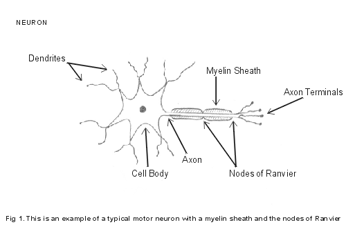
activity. The neuron basically consists of three main components (see Figure
1) First: there is the cell body which contains the cell nucleus and other components
that are necessary for neuron survival and function. Second: there are numerous
dendrites which branch out in order to be able to receive impulses from various
sources. And finally: there is the axon which sends the nerve impulse message
onward to either nervous or muscular tissue. The axon is able to stimulate many
cells by branching out into numerous nerve terminals. Some axons are insulated
by a sheath of myelin, a fatty protein, that enhances the speed of transmission
of the impulse. There are periodic gaps in the myelin sheath which relate to
the cells that form the myelin. These gaps are known as the nodes of Ranvier.
The significance of all this will be revealed below.
The Nervous System
The human nervous system is divided into two major divisions. The central
nervous system (CNS) consists of the brain and the spinal cord. The brain contains
the higher nervous functions such as learning, memory, judgment and language.
It coordinates all sensory and motor activity and it also controls many basic
functions such as respiration, blood pressure, blood flow, heart rate, alertness
and the sleep/wake cycle. All other remaining nervous tissue is collectively
known as the peripheral nervous system (PNS). This system includes the spinal
nerves that exit the spinal cord which branch out to form nerve trunks and then
spread out to form smaller nerves which extend to the very external surfaces,
(the periphery), of the body.
Within most large nerves in the peripheral nervous system there exists three types of neurons. One is a sensory neuron, that is sending messages from the periphery to the spinal cord which will then go up to the brain to tell it what the body is sensing in the way of touch, temperature, pain, vibration and joint position. There are sensory receptors strewn throughout the skin, the muscles, the tendons, and the joints, that allow our nervous system to keep track of the world around us and what is going on in the body. In addition, we have special sensory organs, like the eye and the ear, that allow us to see and hear a limited amount of the light spectrum, and air wave vibration, respectively. The ear also houses the semicircular canals which gives the brain information about the body's location and movement in space.
The second type of neuron in the peripheral nervous system is the motor neuron. After the motor strip in the brain sends a message down the spinal cord, it's the motor neuron that passes it on to muscle groups in the periphery telling them what or what not to do. The instructions may be to either contract and move something within the body, or relax to let complementary muscles do their job. Finally, there is a third type of neuron in peripheral nerves which controls automatic reflexes in the body. These are reflexes that are out of our control but which help us survive in a stressful situation, the so-called "flight or fight" response. How does this all work in practical terms? Here's an example.
Imagine that you are walking down an alley at dusk and someone suddenly grabs your right arm in order to rob you. But with great effort, you manage to wrench yourself away. Here's what would have happened in your nervous system within that fraction of a second. The pressure and pain sensors in your right arm would have sent nerve impulses to the spinal cord which would have crossed over to the left side of your brain to tell you that someone was painfully grabbing your right arm. In response to this insult, your brain would start sending urgent messages from various motor regions instructing the muscles to wrench yourself away from your attacker. At the same time, the photons of light bouncing off your assailant would cause a photochemical reaction in your retina that would result in messages being sent to the visual center of your brain where an image could be developed for your memory. And finally, your autonomic system would be simultaneously engaged in order to increase your respiration, heart rate, blood flow and muscle strength while at the same time making your mouth dry as cotton and your hands sweaty, so that you could achieve your escape. All this in a fraction of a second!!
So you can see that the body's nervous system and its ability to function properly is dependent on the complicated organization of individual parts such as: sensory receptors, sensory, motor and autonomic neurons, the spinal cord, functioning muscle tissue, and the brain. This complex system gives us the ability to detect physical phenomena and to interact and survive within our environment.
Without even considering the biomolecular basis underlying how these messages are sent throughout the nervous system, one can't help but to wonder how each component, listed above, came into existence, one step at a time, as macroevolution dictates, allowing for such a complex and coordinated effort to allow for survival. Medical textbooks are full of life-threatening neuromuscular conditions that result from there being a dysfunction of any one of these vital components that are necessary for life. Conditions such as: peripheral neuropathies that often cause loss of sensory and autonomic function: spinal cord injuries and degeneration: motor neuron diseases such as Lou Gehrig's Disease or muscle diseases such as muscular dystrophy: and numerous degenerative brain conditions such as Alzheimer's and Parkinson's Diseases.
Just try to imagine how a multi-system organism with a complex body plan, absent any of the vital senses that we require, an intact neuromuscular system, or the coordinating activity of the brain, could have survived and been able to propagate. We'll be looking at coordinated sensory and motor function in more detail in future columns. But I just want you to understand how incredibly complex neuromuscular function is and how important it is for our survival. Now onto the nitty gritty of nerve function.
But How Does it all Really Work?
There are three things to consider when looking at how neurons work. The
first is to wonder how the nerve impulse is initiated. In the scenario already
mentioned, where you are walking down a dark alley, it's the sensory receptors
in the skin which detect pain, touch and pressure, that will start the message
toward the brain. And then the cells in the motor strip of the brain will start
the messages back down to the muscles in order to get you to pull yourself away.
Similarly, when light shines through the cornea to the retina, this will ultimately
generate a message that will be interpreted in the brain as the image of your
assailant. Future columns will explain how various sensory organs are able to
convert physical forces such as pressure, temperature, photons of light, and
sound waves, into messages that the brain can interpret. So I won't deal with
this here.
The second question to be asked is how the neuron is able to relay the impulse from where it has been generated to where it needs to be interpreted and have an effect, like in muscle tissue. Alluding to the alley scenario again, when the sensory organs that detect pressure and pain generate nerve impulses they have to be able to travel along the projection of the neuron called the axon (see Figure 1). Similarly, when the brain cells in the motor strip generate messages that are destined to affect muscle activity in the arms and legs, the initial impulse for a given neuron needs to be able to travel along its axon to achieve that function. Exactly how does this happen? That will be the focus of this column.
Assuming that we can understand how the nerve impulse is generated (as future columns will demonstrate) and, secondly, how the once generated impulse can be propagated down the axon of the neuron, it would seem that the third and final piece of the puzzle would involve trying to understand how the neuron is then able to pass on this message to other neurons or muscle tissue. In our alley scenario mentioned above, once the pain and pressure receptors generate a nerve impulse along the neurons, those messages need to be passed on to several other neurons on their way to the sensory strip of the brain. Like-wise, when the neurons in the motor strip generate the messages that are needed to activate your muscles, those impulses need to be passed on to several other neurons until they finally stimulate muscle cells to cause the desired action. Without the ability for the neuron to pass on the desired message to the right tissue, the ability to generate the message and allow it to travel down the axon would be useless. Next month's column will explore this question.
Basic Knowledge in order to understand Nerve Conduction
In order to be able to appreciate the complexity of the neuron's ability
to conduct an electrical impulse, it is important that you understand five main
concepts about the cell, its plasma membrane, and the resulting relationship
between the chemicals inside and outside of the cell.
1. The chemical composition inside the cell (intracellular) is vastly different than the chemical composition outside the cell (extracellular). (see Table 1) The predominant positively charged ions (cations) in the cell are potassium (K+) with much lesser amounts of sodium (Na+). And the predominant negatively charged ions (anions) in the cell consist largely of phosphate-containing biomolecules and proteins, all of which are necessary for cell function and survival. The main cations outside the cell are sodium (Na+) with much lesser amounts of potassium (K+). And the predominant anions outside the cell are chloride (Cl-). Separating these two competing chemical solutions is the plasma membrane of the cell.
|
Chemical
Composition of ECF and ICF
|
|
|
ExtraCellular Fluid |
IntraCellular Fluid |
| Sodium Ions (+) 143 units Potassium Ions (+) 5 units Chloride Ions (-) 117 units |
Potassium Ions (+)
157 units Sodium Ions (+) 14 units Phosphate Ions (-) 113 units |
| Bicarbonate (-) 27 units |
Protein bases (-) 72 units |
Table 1. Note the difference in the relative concentrations of sodium and potassium ions within the ECF and ICF.
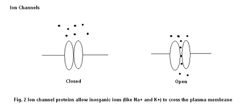
2. There is a tendency for each positive ion (Na+, K+) to try to cross the membrane barrier down its concentration gradient. In other words, given the chance, potassium is likely to try to come out of the intracellular fluid and into the extracellular fluid because its concentration in the cell is much higher than outside the cell. Similarly, sodium, if given the opportunity, is likely to try to enter the cell from the extracellular fluid because its concentration is much higher outside the cell than inside it. The cell membrane contains certain proteins that act as ion channels for specific ions (see Figure 2). So there exists potassium and sodium ion channels. In addition, depending on the cell type, each ion channel has a different degree of efficiency with regard to allowing its specific ion to move along its concentration gradient (remember, if their respective ion channels open up, sodium wants to go into the cell and potassium wants to go out). Therefore, the plasma membrane has a variable permeability to each of these ions which is dependent on how many of the ion channels are open at a particular time. In fact, depending on the cell type, the plasma membrane is between 10 to 100 times more likely to let potassium out of the cell than let sodium in.
3. All cells have an energy-dependent sodium-potassium pump in the plasma membrane which pushes three sodium ions out of the cell for every two potassium ions that it pumps back in. The net pressure that exists for water to move between two solutions separated by a semi-permeable membrane is dependent on the total amount of particles within each solution that are incapable of transferring between them. The fluid outside the cell has a lot of inorganic particles (Na+, K+, Cl-) which are offset in the cell by some inorganic particles (K+, Na+) and organic particles, such as phosphate-containing biomolecules that are used for energy transfer and structural and enzymatic proteins. But remember that by way of the ion channels, the inorganic particles are actually capable of passing through the membrane while the organic ones can't. Over time this can result in the total concentrations of the inorganic particles inside and outside of the cell becoming equal while leaving the organic particles inside the cell to attract water. This will make water from outside the cell go into the cell and cause expansion and ultimately death by explosion. In order for the sodium-potassium pump to push sodium ions out of the cell and bring potassium ions in against their respective gradients, it requires energy. By pushing out more sodium ions than allowing potassium ions back into the cell, the sodium-potassium pump is able to maintain the proper size of the cell. When the cell runs out of energy, such as occurs with low sugar and lack of oxygen, the sodium-potassium pump fails, water comes into the cell, and it expands, explodes, and dies. In fact this is the final common pathway for cell death, just as cardiopulmonary arrest is the final common pathway for bodily death.
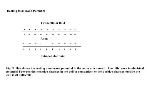
4. The sodium-potassium pump helps maintain the proper amount of potassium in the cell. However, in all cells, the potassium ions have a tendency to leak out by way of their ion channels in the order of 10 to 100 times easier than sodium ions. Because of this, there is a build-up of a negative charge within the cell as compared to outside the cell because more K+ ions have gone out of the cell compared to Na+ ions going back in. This gives rise to what is called the membrane potential. However, in addition to this, now that the inside of the cell has a negative charge with respect to the outside of the cell, there is a tendency for the K+ ions to go back inside the cell to counterbalance this negativity. In other words, the K+ ions have a tendency to travel down their concentration gradient out of the cell and do it more efficiently than Na+ ions which results in a negative membrane potential which then attracts the K+ ions back into the cell. The final balancing act results in what is called the resting membrane potential, which, depending on the cell type usually ranges from -40 to -90 mV (millivolts). The resting membrane potential for a neuron is about -70mV. (see Figure 3)
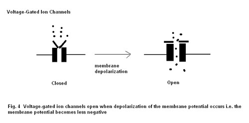
5. It may be evident to you by now that the ability for the neuron to produce this resting membrane potential is largely dependent on the ion channels which results in a variable plasma membrane permeability for Na+ and K+ ions. It turns out that ion channel activity can be regulated by way of either membrane voltage changes, so called voltage-gated ion channels, (see Figure 4), or by chemical messengers that lock onto specific sites that activate the ion channel, so called ligand-gated ion channels. (see Figure 5). The ligand-gated ion channels will become important to our discussion in next month's column when we look at the mechanism by which the neuron is able to transfer its message and affect the function of another nerve cell or a muscle cell To recap: when the membrane potential becomes less negative, specific ion channels will open, allowing ions to transfer between the intracellular and extracellular fluid which directly results in a further change in the membrane potential. (remember, K+ wants to go out of the cell and Na+ wants to go into the cell).

The Action Potential of the Neuron
If you've digested these five principles then you're ready to dive in and
understand how the axon of the neuron is able to conduct an electrical impulse
on its way to transmitting it to another neuron or muscle cell. Here goes!!
Neurons and muscle cells are excitable cells in that when they are properly stimulated, they either transmit an impulse, as in the neuron, or contract, as in the myocyte. Both of these cells are capable of their respective actions because they each have the capability of totally depolarizing and repolarizing their membrane potentials. That is, when properly stimulated, the neuron will change its polarity, going from - 70mV to +40 mV, and then it will return to its original resting membrane potential of -70mV again. Here's how.
When the axon of a neuron is adequately stimulated, it will begin to open some of its sodium ion channels which will allow the positively charged Na+ ions to enter the cell. As this occurs the negative resting membrane potential of -70mV begins to become less negative because positive sodium ions are entering the cell. If the stimulus is strong enough, this will begin to open more and more voltage-dependent sodium ion channels, thereby allowing a maximal amount of Na+ into the cell. So much so, that the membrane potential will now become positive, usually in the range of +40 mV. The membrane potential will have depolarized by going from being at first negative, to now being positive. This is called the generation of the action potential.
However, if this change in the membrane potential were to remain as a constant, i.e. if the resting membrane potential were to stay at +40 mV after a neuron was stimulated for the first time, this would render the neuron as essentially useless for ongoing communication to other parts of the neuromuscular system. It would be like having to replace the wires in your TV after every time you used it. So it's just as important to understand how the neuron can go back to its previous resting state and be prepared for another message as it is to understand how it can generate an action potential.
The process by which the neuron is able to achieve its original resting membrane potential of -70mV is called repolarization. What happens is that as sodium floods into the axon, causing the membrane potential to reverse its polarity and become + 40 mV, this causes the voltage-gated sodium ion channels to start to shut down to stem the tide. At the same time, voltage-gated potassium (K+) channels open up, allowing K+ to escape from the cell, thereby making the membrane potential dip back down towards its original level. As the membrane potential approaches its resting value of -70mV the voltage-gated K+ channels close down to their resting state and the sodium ion channels revert to their closed state in order to maintain the resting membrane potential and allow the neuron to be prepared for another message.
A quick look at what has just happened when a neuron produces an action potential will make you realize that after the repolarization there would appear to be more Na+ ions and less K+ ions in the cell than there were before the action potential took place. Remember, the membrane depolarized because of the entry of Na+ ions into the neuron and it repolarized, not because the extra Na+ ions left, but because K+ ions, which are also positive, left the neuron to make the membrane potential return to -70mV. The sodium-potassium pump, by pushing sodium out of the cell and bringing in potassium against their gradients makes the necessary correction in order to maintain neuron function.
But how does the action potential travel down the axon? As the region of the axon that is being stimulated becomes totally depolarized, going from an initial resting membrane potential of -70mV to +40 mV, this change affects the voltage-gated sodium channels that are right next door. This causes the neighboring membrane to also let in more and more sodium as more and more sodium ion channels open up. The process then continues along the axon until it reaches its numerous axon terminals. (see Figures 1 and 3)
One differentiating feature among neurons is the presence or absence of myelin around the axon. In general, without myelin, the speed at which the nerve impulse is conducted along the axon is slower in comparison to an axon encased in it. This is because, in the presence of insulating myelin, the action potential cannot travel directly along the axon but must literally jump between the gaps in the myelin where the extracellular fluid is in direct contact with the axon membrane. These gaps are called the nodes of Ranvier and the jumping action is known as saltatory conduction. (see Figure 1)
On a practical level, the nervous system consists of different sized, unmyelinated, and variably myelinated neurons, each with different impulse velocities. The larger diameter a neuron is, the faster it will send an impulse. Therefore, the fastest conducting nerves in the body are the largest and most myelinated nerves. This just so happens to coincide with the ones that have a direct effect on motor function. Right behind them are the nerves for cutaneous sensation. And the pain fibers are the slowest of all. That's why when you are injured, you'll feel the insult right away but there will be a delay in your feeling the deep pain associated with the injury.
Now, think about that episode in the dark alley with your assailant. It's important for your survival that the sensory nerves be able to not only detect the hands of your assailant on your arm, but also to be able to send a quick message to your brain telling you of this state of affairs. It's not really important for you to be aware of the pain that you will soon feel. In fact it may be counterproductive and interfere with your defensive maneuvering. At the same time, your eyes and ears will be sending messages to your brain as well. But don't forget, these organs are not very far from the brain as compared to your arms and legs. So the speed at which their messages travel along the optic and auditory nerves may not necessarily have to be as fast as what is needed for your escape.
Finally, your brain will be sending messages to coordinate many of your muscles into a concerted action designed to evade your attacker and get to safety. You can see why it is so very important that the nerves that send the fastest messages should be the ones that allow for evasive action. Sensory nerve speed is important as well, but it doesn't necessarily need to be as fast as the motor nerves and with good reason. For myelin consists of fatty protein which requires a lot of energy from the body for formation.
There's a hierarchy within the body of what is needed for defense and survival. It's no benefit to the body to use up too much energy by providing a lot of myelin for neurons that don't necessarily provide information that is needed on an immediate basis, such as for deep pain. And you have to decide which neurons are going to be your ace in the hole when the going gets tough. Being aware of danger is very important for survival, but being able to elude danger requires more finesse and energy. The body's nervous system seems to be inherently designed for efficient energy preservation while at the same time allowing for an adequate functional capacity for survival.
Questions for Macroevolution
There are many reasons why neurons do not work properly resulting in neurological
dysfunction. This would have spelled death for a multi-system organism with
a complicated body plan, no matter where it was in its development. The neuron
must be able to be stimulated, allow the impulse to travel along its axon in
a timely manner, and be able to pass on the message to its intended target;
be it either another neuron or a muscle cell. Regarding the topics that have
been covered in this column, there are several questions that would seem to
need to be answered before one could suppose, or should propose, that this system
developed by the random forces of nature, one step at a time, while still remaining
functional to allow for survival.
It is evident that the presence of voltage-regulated ion channels that switch on and off at the right times to allow for the controlled depolarization and repolarization of the neuron is vital for neuromuscular function. So one must wonder about the step by step development of neuron function with respect to the presence or absence of both the Na+ and K+ ion channels and their ability to be properly voltage regulated.
Which came first, the Na+ ion channels or the K+ ion channels? When did they each become voltage-gated? How is it that the voltage regulation allows for the proper sequencing of ion motion in a way that results in the neuron being able to repetitively pass along impulses of information? If all of this developed one step at a time, then wouldn't one have to at least postulate how the neuron functioned during these intermediate phases of development in which one or more of the components that we know are necessary for it to work were absent?
Where does the sodium-potassium pump fit in to all of this? We have seen that not only is the sodium-potassium pump necessary for all cells to survive, it is also vital for the nerve cell to be able to repetitively function. And without neuromuscular function, how could a multi-system organism with a complex body plan survive?
The Pathophysiology behind one class of Neuron Dysfunction
Those of you who have been reading my columns have probably noticed that
I inevitably use examples of human disease to illustrate my points about what
can happen when one small part of a complicated system doesn't work right. I
didn't want to disappoint you so I will briefly discuss here one of the commonest
classes of neurological conditions that can afflict humans.
These are the demyelinating disorders. As the name implies; the demyelinating disorders are neurological conditions in which the myelin, that surrounds the nerves that conduct quick impulses in order for our neuromuscular system to work efficiently, is defective in some way. I think that you'll agree that any multi-system organism with a complex body plan like our own, that had defects as described below would have great difficulty in winning the battle for survival. Some of the more common conditions are:
Multiple Sclerosis (M.S.): A chronic, relapsing disorder involving the brain, the optic nerves and the spinal cord, in which areas of inflammation attack and injure the myelin around these nerves. Symptoms are related to the nerves that are involved, such as: double vision, blindness, slurred speech, weakness in arms and legs with inability to feed oneself or walk, inability to sit, inability to control the bladder, inability to swallow safely, confusion, depression and emotional lability
Guillain Barre Syndrome (Acute Inflammatory Demyelinating Polyneuropathy): A condition, which often follows an acute infection, in which inflammation of the peripheral nerves results in the injury and dysfunction of the myelin sheath surrounding the axons. Typical symptoms include: numbness and weakness of the legs resulting in the inability to stand and walk, followed often by quick progression up the body to the arms, difficulty swallowing, and it can become life-threatening if it involves the nerves to the muscles of respiration because then you can't breathe effectively.
Charcot-Marie-Tooth Disease: This is a group of inherited disorders that involve an abnormality in a specific protein that is found in the myelin sheath. The resulting abnormality usually involves an excessive amount of abnormal myelin surrounding the neuron. This often results in various neuromuscular abnormalities and dysfunction, ranging from foot and ankle weakness and deformities with poor overall mobility, to severe motor-sensory dysfunction affecting the entire body, manifesting symptoms that are similar to M.S. and G.B.S. as described above.
So one can readily see that myelin is very important for overall neuromuscular function in the human body and thereby also, survival on earth. Therefore, given this information, one could further ask those who support the mechanics of macroevolution to explain the development of life: How did a multi-system organism with a complex body plan develop a nervous system that included myelin in all the right places? How did it survive without it? And where does myelin fit into the scheme of things that includes the sodium-potassium pump and the different voltage-gated ion channels already mentioned above? These are only a few of the questions that should require logical and definitive answers before macroevolution should be considered a truth of science. For as in my prior columns, I have only scratched the surface of this indeed very complex bodily function.
We've Only Just Begun (Wasn't that the name of a song?)
But what happens when the action potential finally reaches the axon nerve
terminals? How is it able to transmit the impulse on to other nerve or muscle
cells? And how does that impulse affect the nerve or muscle cell on the receiving
end? The answers to these questions and the further questions that these answers
engender for macroevolution will be dealt with in next month's column. Please
join me then in: Wired for Much More than Sound---Neurons and how they work-Part
II-- The Transfer
Dr. G.
Howard Glicksman M. D. graduated from the University of Toronto in 1978. He practiced primary care medicine for almost 25 yrs in Oakville, Ontario and Spring Hill, Florida. He recently left his private practice and has started to practice palliative medicine for a Hospice organization in his community. He has a special interest in how the ethos of our culture has been influenced by modern science’s understanding and promotion of what it means to be a human being.
Copyright 2004 Dr. Howard Glicksman. All rights reserved. International
copyright secured.
File Date: 7.01.04